On February 1, 2020, a fascinating new exhibition that explores the intersection of art and science opens in the newly renovated and enlarged Science Gallery at the Bruce Museum in Greenwich, CT.
“Under the Skin” highlights a dozen recent scientific discoveries through a combination of stunning imagery and real biological specimens.
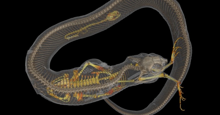
Hog-Nosed Snake with Prey, CT Scan. Image courtesy of Dr. Ed Stanley and Dr. David Blackburn
The exhibition, on view through July 19, 2020, showcases images made possible by a remarkable array of technologies – CT scanning, infrared and UV imaging, scanning electron microscopy, and more – that reveals the extraordinary beauty of nature that often lies just below the surface. All of the images presented in the exhibition, from a section of a dinosaur bone photographed in cross-polarized light, to a CT scan of a hog-nosed snake engorged with prey, were captured in the last five years, thus representing the cutting edge of modern imaging.
Pelicans, Thermal Image. Image courtesy of Dr. L. Witmer, Dr. R. Porter, and Dr. G. Tattersall
A number of the images in “Under the Skin” highlight new scientific discoveries that were undreamt of a decade ago. Specimens from the Bruce Museum Collection and on loan from other collections complement each image and reinforce the role of museums as stewards of natural history.
“Nature is full of beauty, at scales great and small,” says Dr. Daniel Ksepka, Curator of Science. “While each represents a research breakthrough, these striking and, in many cases, prize-winning images can be considered art in their own right.”
Roosterfish, Cleared and Stained Specimen. Image courtesy of Dr. Matthew Girard
Visitors will peer into the inner ear of a tiny frog, marvel at a chameleon whose bones glow in UV light right through its skin, and learn how we can trace the growth rate of a 10-ton dinosaur from microscopic structures in its bones. Exploring the relationship between light and nature, visitors will discover that flying squirrels glow a fluorescent pink in UV light, pelican pouches burst into color when viewed with infrared imaging, and fossil cells transmit a kaleidoscope of color under polarized light.
A dozen images highlighted in the exhibition have been especially printed on metal to provide more detail and depth. “It’s taking the images to the next level, giving them a polished, almost luscious look,” says Anne von Stuelpnagel, Director of Exhibitions. “The photos themselves are from the researchers, and they deserve credit for taking photographs of such tiny or intricate things and at such high resolution. We wanted to do them justice.”
2Kviews
Share on Facebook
 Dark Mode

 No fees, cancel anytime


15
0