19 Groundbreaking Images By This Photographer Offering A Glimpse Into The Miracle Of Birth
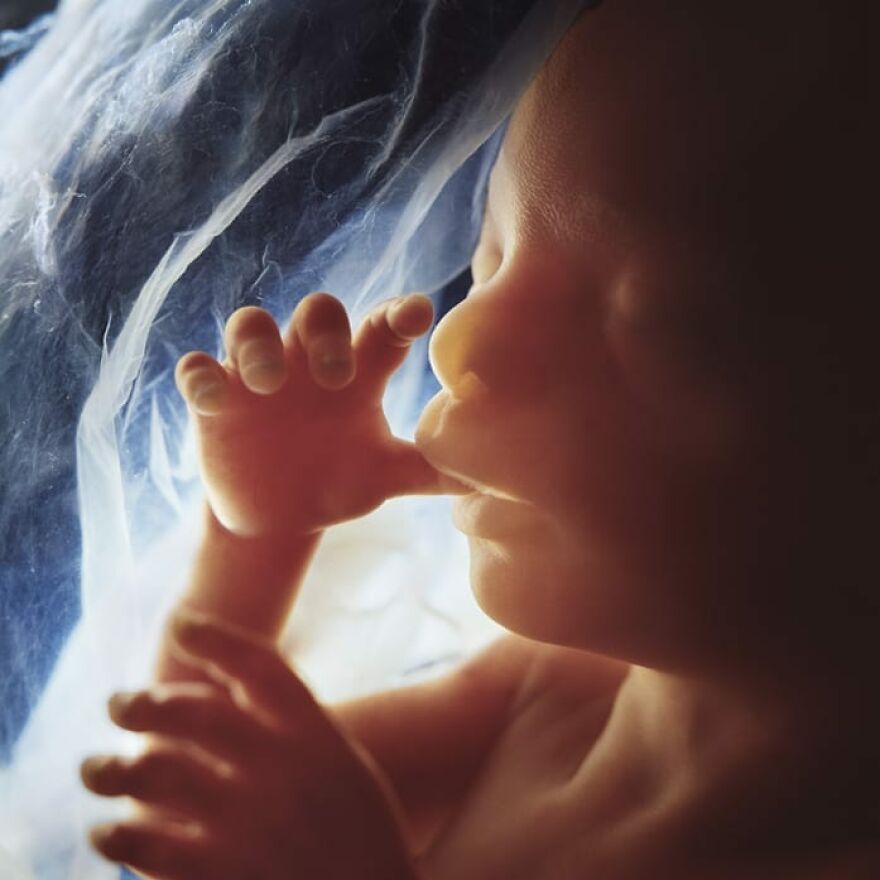
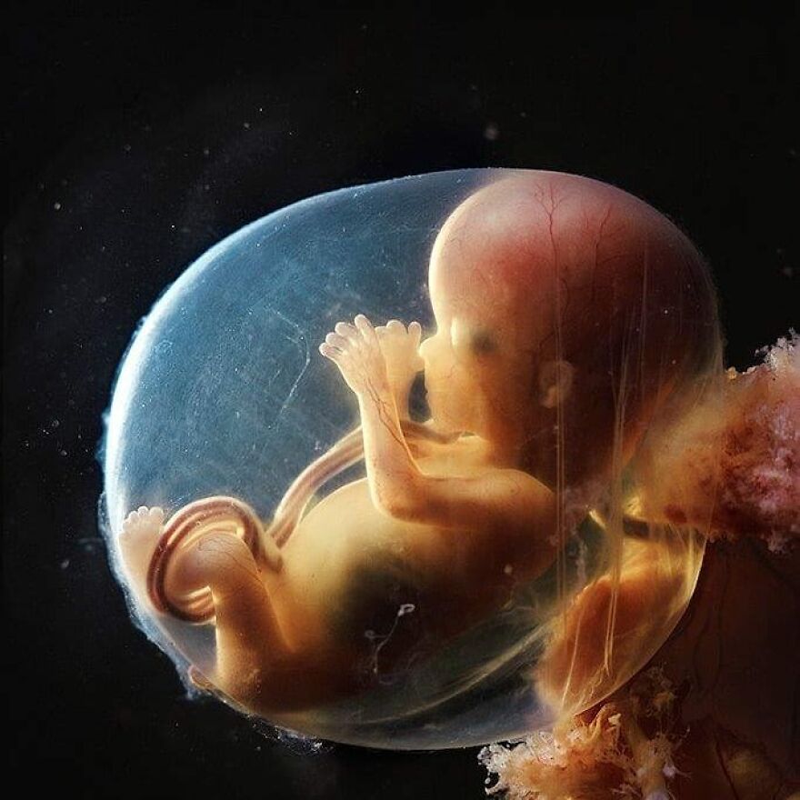
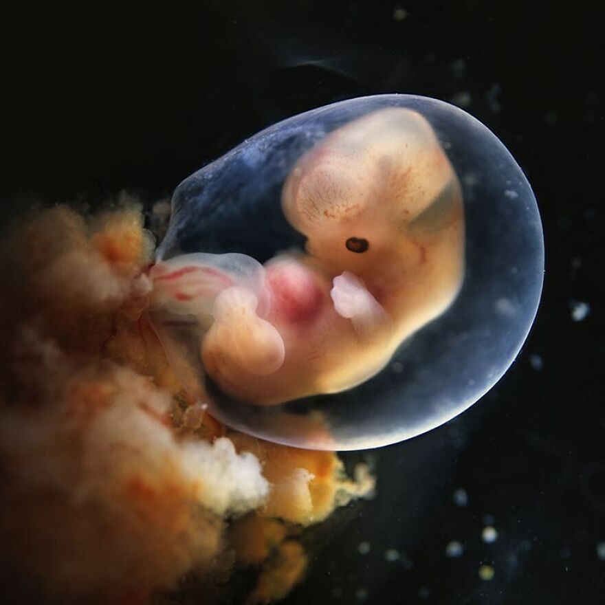
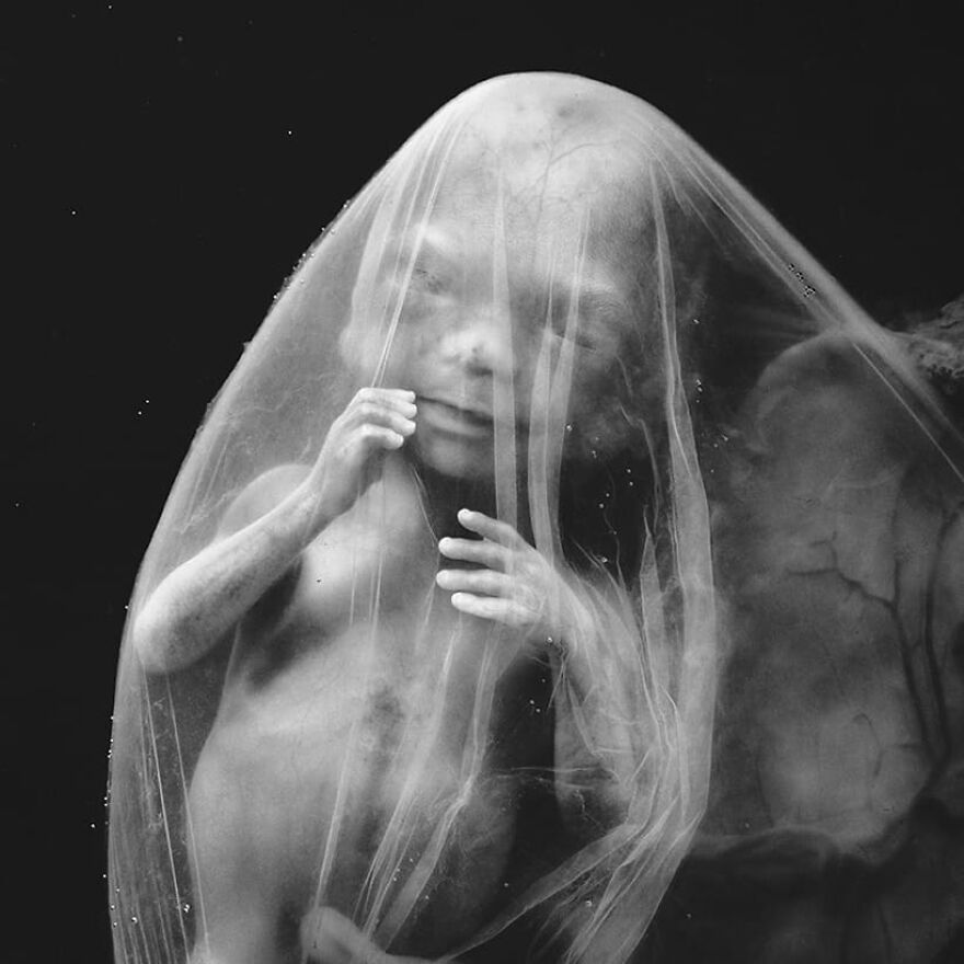
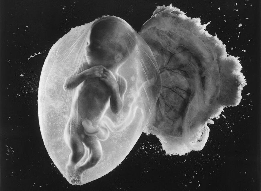
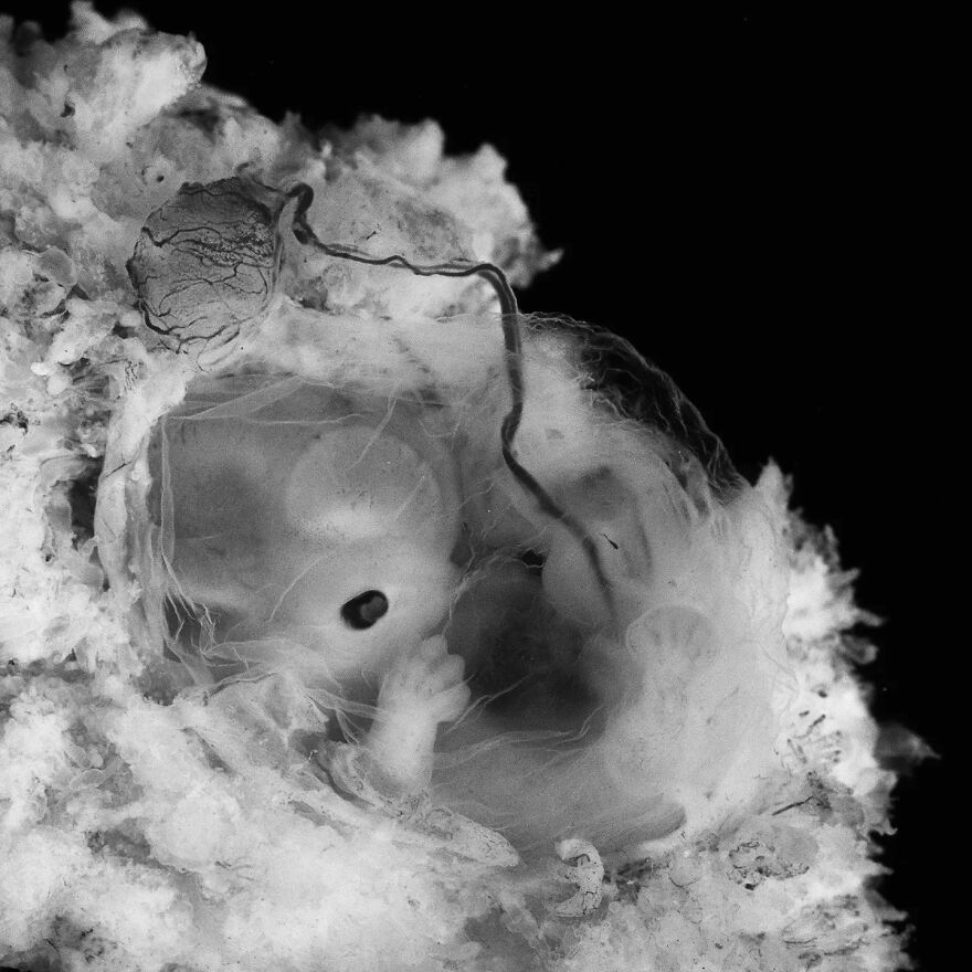
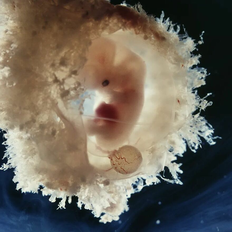
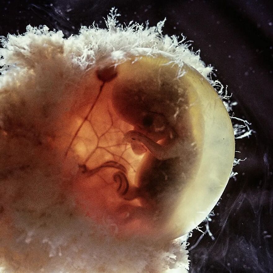
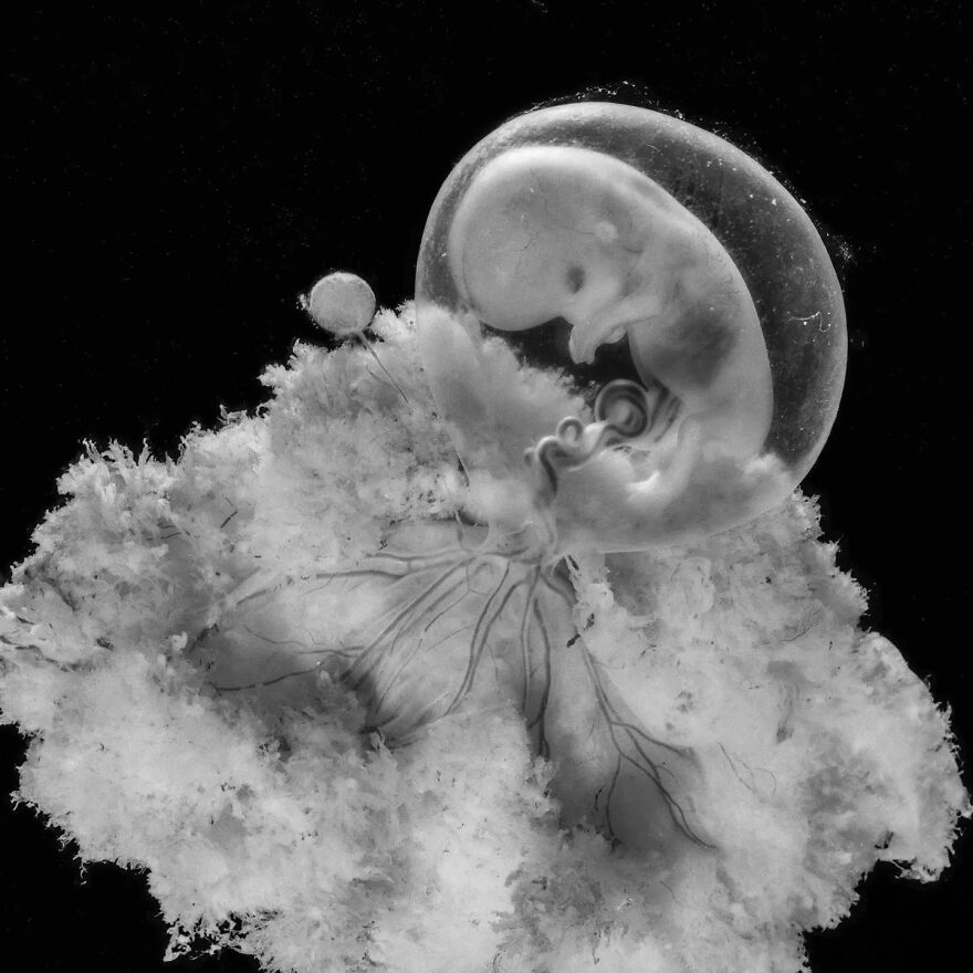
InterviewIn April 1965, Life magazine unveiled "Foetus 18 Weeks," a photograph by Swedish photojournalist Lennart Nilsson. This image, depicting a fetus in its amniotic sac, marked the beginning of Nilsson's research on prenatal development. The photographer collaborated with Professor Axel Ingelman-Sundberg and utilized innovative endoscopic technology, with which he was able to capture groundbreaking images of unborn children.
Today, Nilsson's work still resonates with audiences worldwide. The images captured by him offer a glimpse into the miracle of birth. Scroll down to view a selection of the Swedish photographer's finest shots, and to learn more about his story with insights from Anne Fjellström, the curator of the Instagram account dedicated to Lennart’s work, with whom we had the pleasure of speaking.
More info: Instagram | lennartnilsson.com
This post may include affiliate links.
Fetus, 20 weeks, 1974
I still have the first edition of his book "How Life Begins." This was the cover photo.
Bored Panda reached out to Anne Fjellström, Nilsson’s stepdaughter, who now curates the Instagram account dedicated to preserving the photographer’s legacy. Firstly, we found out more about the inspiration to curate the social media profile: “We have had the account since 2016. Lennart Nilsson passed away in January 2017. We tried to use Facebook in the beginning but found it much easier to communicate using Instagram. It was a complement to our website with the aim of spreading Lennart Nilsson's life's work and maintaining interest in his photographic works. We want his images and films to act as a source of knowledge and inspiration, and give people a greater understanding of themselves and what is close to them.”
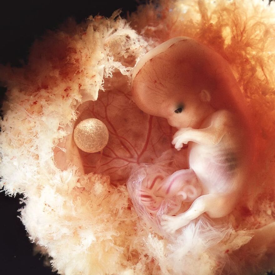
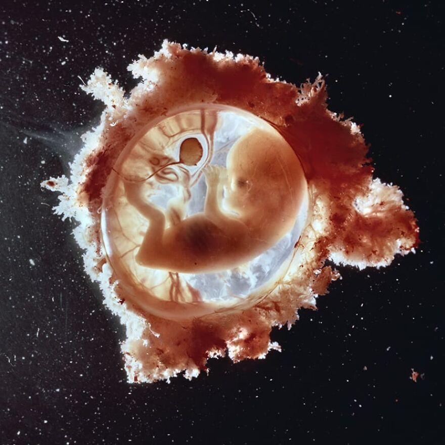
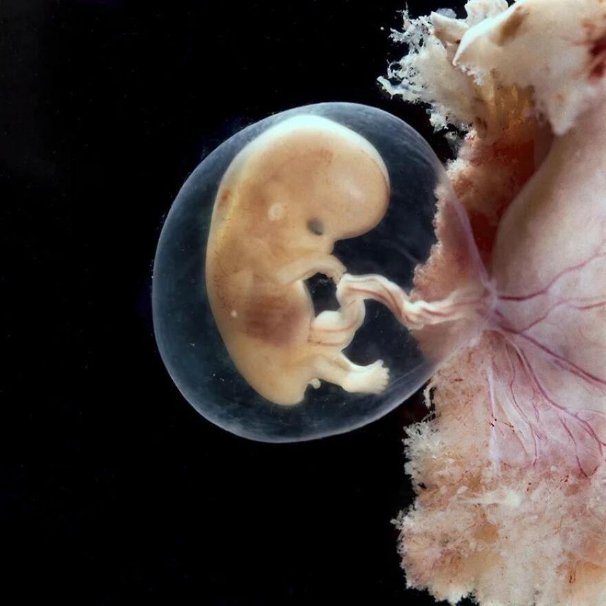
Fetus, 11-12 weeks
About 8 weeks? Just mentally comparing with our son's sonogram. Obv due to the different technology, that's not easy nor fool-proof.
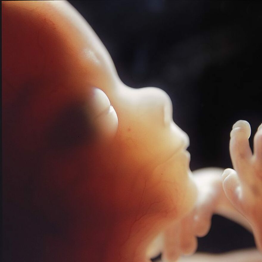
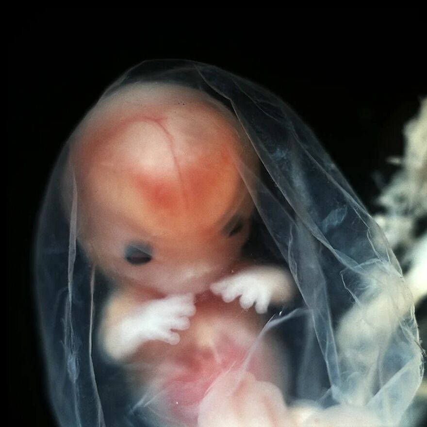
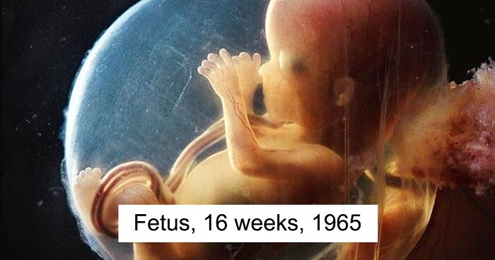
Fetus, 16 weeks, 1965
Lennart Nilsson's photography is renowned for its groundbreaking images of human development and scientific subjects. We were wondering what the process of selecting and curating content for the profile looks like. Fjellström shared with us: “We tried, in the beginning, to show the diversity of his work during his 70 years as a photographer. The response was not as great as when we show the images from his series 'A Child is Born' so we mainly show images from that story. But we are quite restrictive. Not everyone understands the meaning of 'Copyright'. Most people don’t know about his earlier works. That is why we show more on the website to try to communicate his whole life journey as a photojournalist. We might change the content on Instagram further on.”
"Spaceman",
Fetus, 13 weeks, 1965
Anne told us about some of the most memorable responses she’s received from followers of Nilsson’s Instagram profile: “It is mainly the response on the A Child is Born material. We all have such different views, and that is interesting. That material has had an enormous impact and still has. We try to communicate Lennart’s aims with the material, and it is not always what others believe, or want. Most of the images from that series were photographed in the 60s. The world was quite different from today.”
Fetus, 20 weeks, approximately 20 cm
From the series "A Child is Born", 1965
Lastly, we were eager to know what message or impact Fjellström aims to convey to the followers of the photographer's account. Anne said: “That of Lennart. His vision. To make the invisible visible. And to communicate science to the public. Show the greatness in the everyday life. He was a very curious person, and that curiosity was what drove him through his life. And his love for photography. Making it possible to tell us stories with images.”
Fetus, 16 weeks, 1965
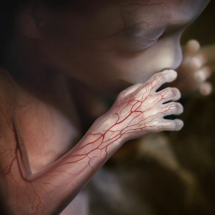
The network of blood vessels for the arm and hand is visible through the thin skin.
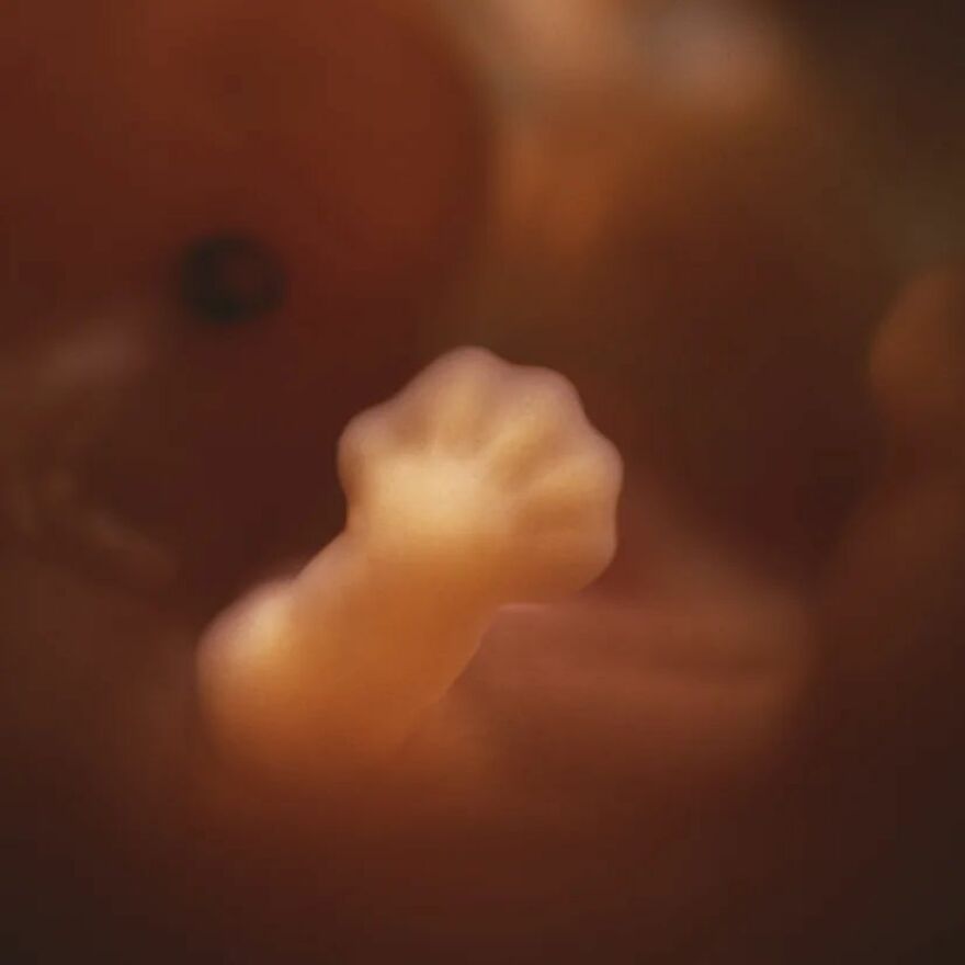
Development of the hand, week 8
From the book "A Child is Born", 2003
"Foetus 18 weeks", 1965
Fetus, 8 weeks (10), approximately 4 cm (1.6 inches)
From the book "A Child is Born", 1965
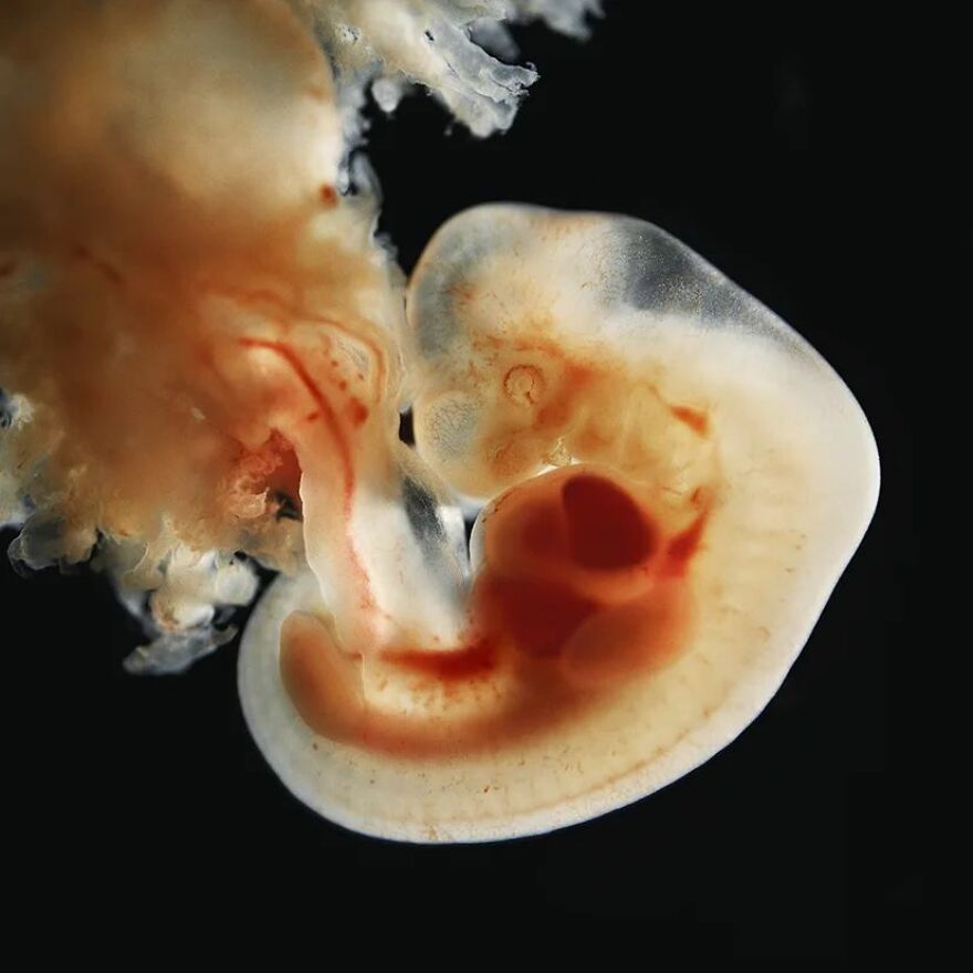
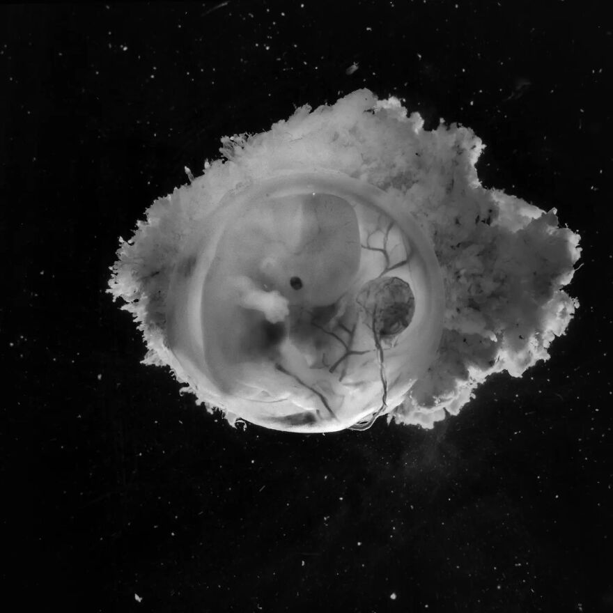
Embryo, 6 weeks, 1,5 cm
From the series "A Child is Born", 1965
I really wanted to enjoy these photos for the amazing photos that they are, but all I could think is some idiot will use these as a reason to be pro-life and continue chipping away at women rights.
Yeah that was my first fear when I saw this, like these are really cool but I feel like these could be easily used to be like “look how developed it is! How could you kill that?” I also wish I knew how exactly these were gotten, I have to think they’re at least a little computer assisted.
Load More Replies...Only one of these images is of a live infant taken with a laparoscopic camera. the others are all deceased. The hospital called him whenever there was a fetus available and that he had to complete his photography within a few hours. He had an aquarium-like tank at the hospital to suspend the deceased fetus in to take the pictures, which is why they look like they are floating.
I really wish these were accurately captioned at what point in development they were because as near as I can tell the earliest any of these were was ~8-10 weeks at the very least.
Thanks for your suggestion! We've just added some captions :)
Load More Replies...I really wanted to enjoy these photos for the amazing photos that they are, but all I could think is some idiot will use these as a reason to be pro-life and continue chipping away at women rights.
Yeah that was my first fear when I saw this, like these are really cool but I feel like these could be easily used to be like “look how developed it is! How could you kill that?” I also wish I knew how exactly these were gotten, I have to think they’re at least a little computer assisted.
Load More Replies...Only one of these images is of a live infant taken with a laparoscopic camera. the others are all deceased. The hospital called him whenever there was a fetus available and that he had to complete his photography within a few hours. He had an aquarium-like tank at the hospital to suspend the deceased fetus in to take the pictures, which is why they look like they are floating.
I really wish these were accurately captioned at what point in development they were because as near as I can tell the earliest any of these were was ~8-10 weeks at the very least.
Thanks for your suggestion! We've just added some captions :)
Load More Replies...
 Dark Mode

 No fees, cancel anytime


