2016 Nikon Macro Photo Contest Winners Show The World Like You’ve Never Seen Before
Nikon has just announced the winners of its annual Small World Photomicrography competition, and as you can see from these stunning photographs, bigger isn't always better.
The competition is in its 42nd year and this year over 2000 people from 70 countries entered. For those that don't know, photomicrography is the practise of taking a photograph through a microscope or similar magnifying device in order to capture the intricate details of things invisible to the human eye. From the proboscis of a butterfly and the foot of a beetle to espresso coffee crystals, the pictures below give us a whole new way of looking at world. The categories are divided into winners, honorable mentions, and images of distinction, and you can find the full list on the Nikon Small World website.
More info: Nikon Small World (h/t: demilked)
This post may include affiliate links.
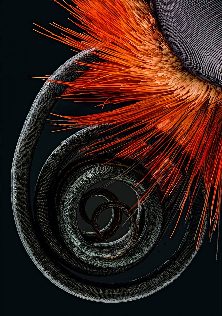
Fourth Place. Butterfly Proboscis
Stunning! One of the best macro shots I've ever seen. Congradulations on derserving win!
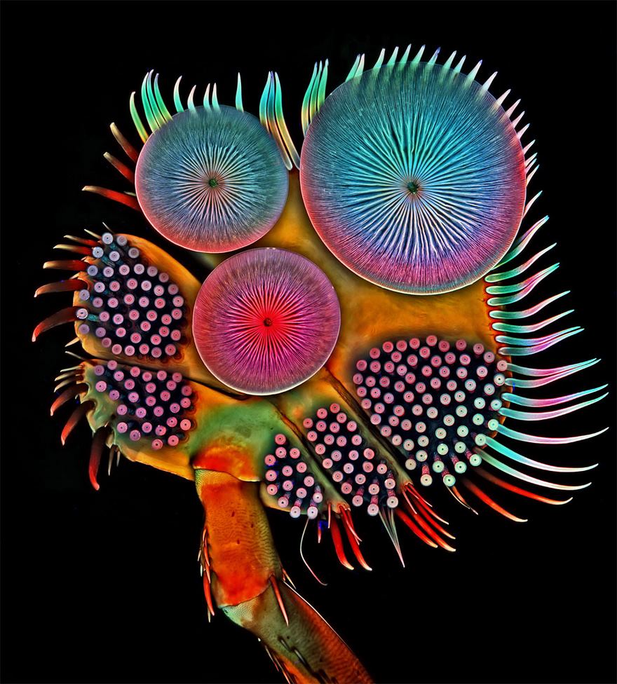
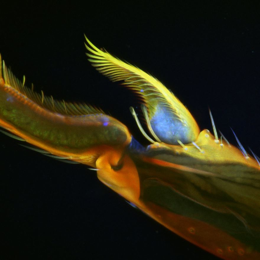
Fifth Place. Front Foot (Tarsus) Of A Male Diving Beetle
Eyes Of A Jumping Spider
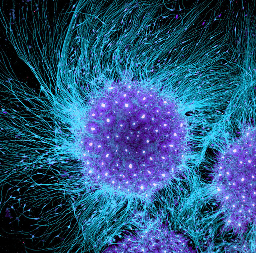
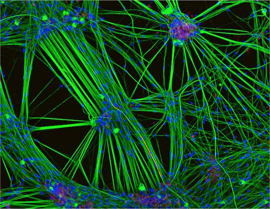
Nineteenth Place. Human Neural Rosette Primordial Brain Cells
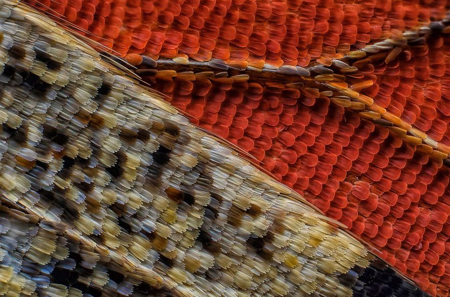
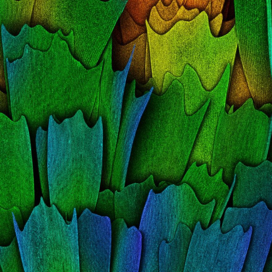
Eleventh Place. Scales Of A Butterfly Wing Underside
All butterfly wings look like they're made of a million little feathers.
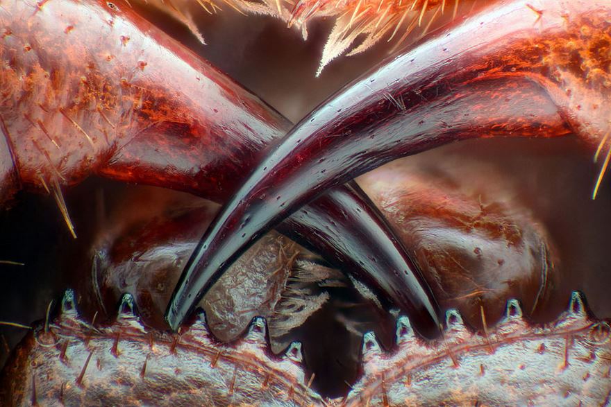
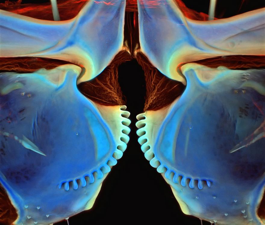
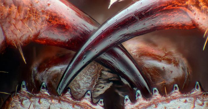
Thirteenth Place. Poison Fangs Of A Centipede
At first glance I thought it was an animals teeth. Either way, I would not want to be bit by them!
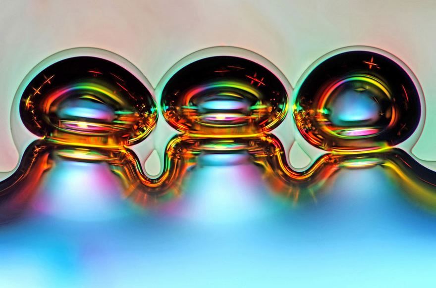
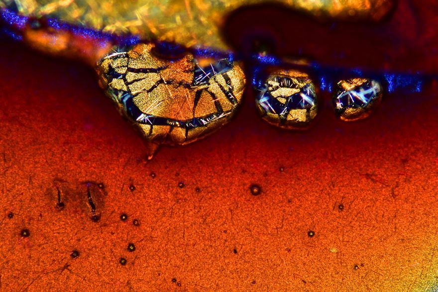
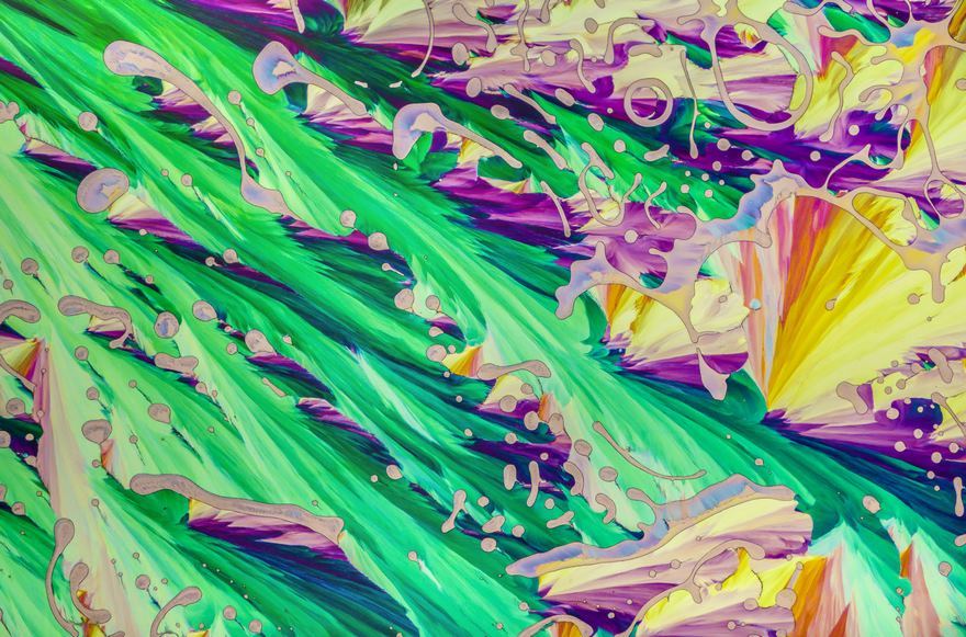
Sixth Place. Air Bubbles Formed From Melted Ascorbic Acid (Vitamin C) Crystals
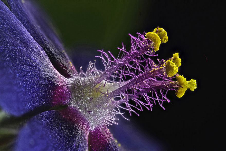
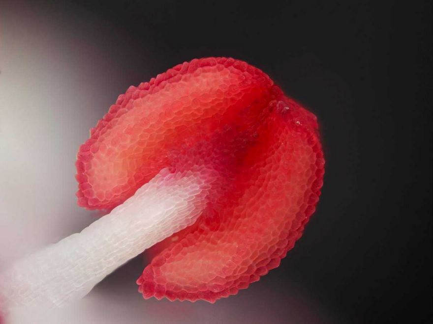
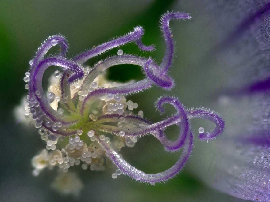
Eighth Place. Wildflower Stamens
This is what the bees and butterflies see as they approach. How enticing.
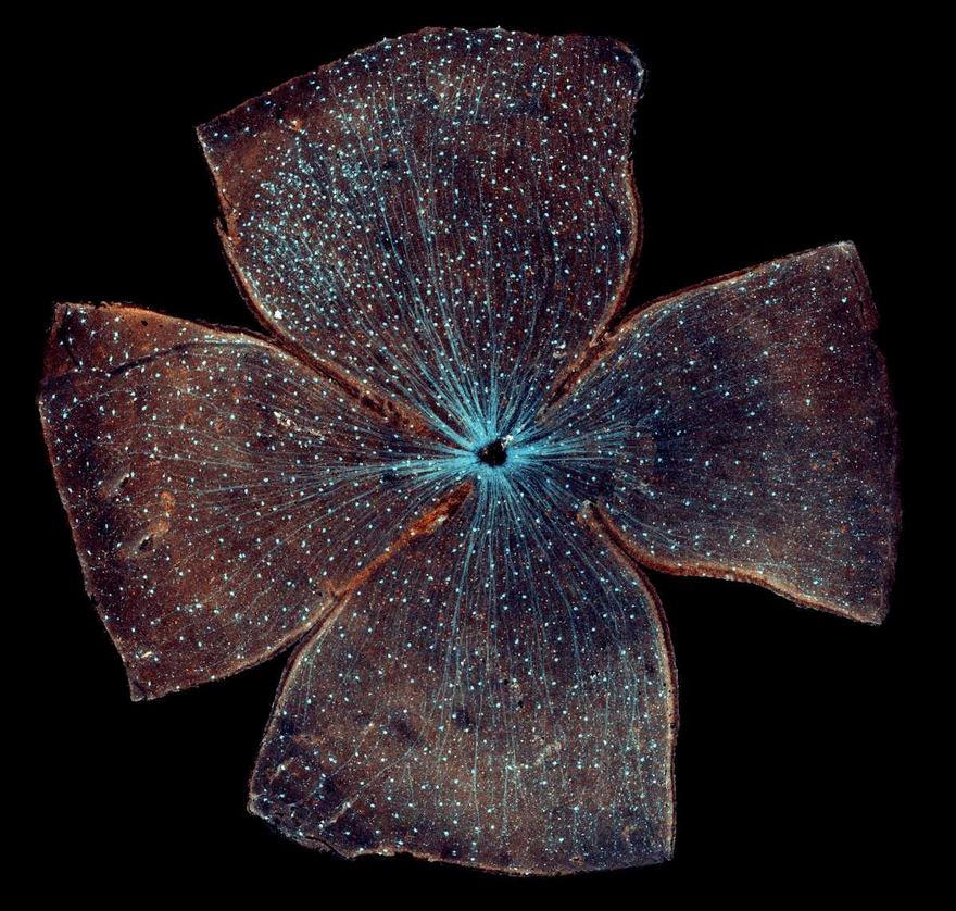
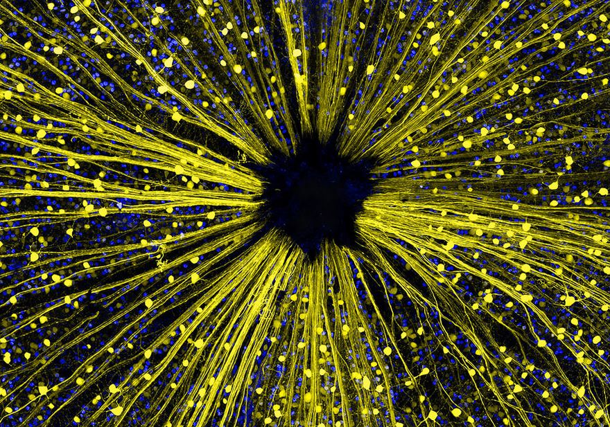
Retinal Ganglion Cells In The Whole-Mounted Mouse Retina
Ninth Place. Espresso Coffee Crystals
That one to the left looks like a snakes head, and you can even see a slithering tongue sticking out!!
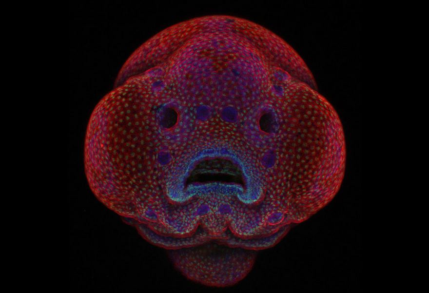
First Place. Four-Day-Old Zebrafish Embryo
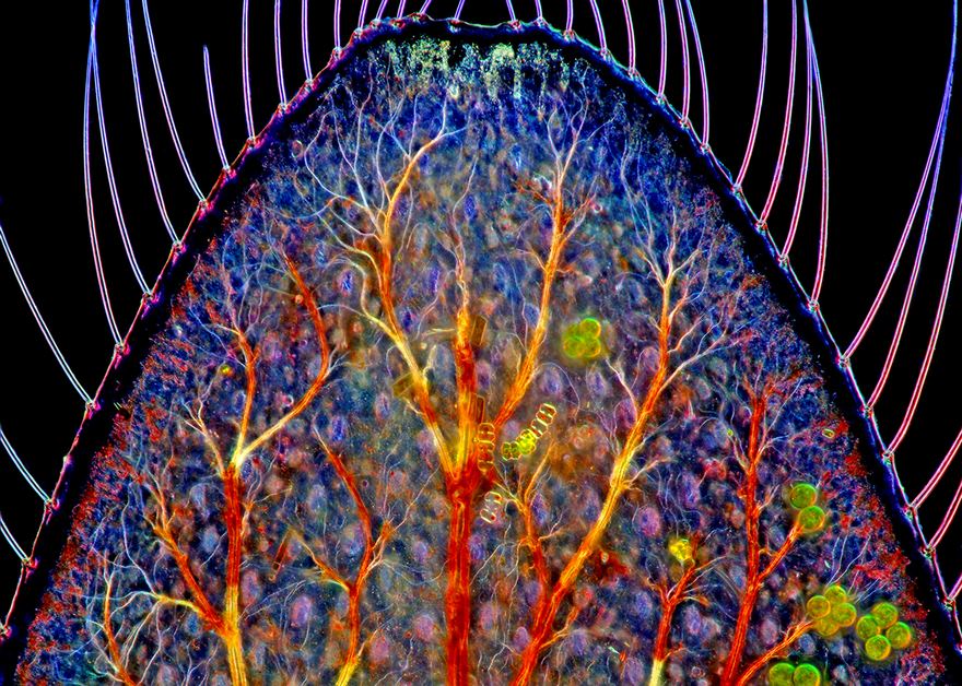
Caudal Gill Of A Dragonfly Larva
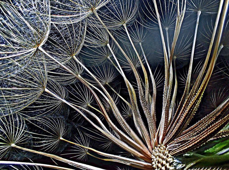
Goatsbeard Flower
Aren't these the same flowers we pick as kids, then make a wish and blow on them?
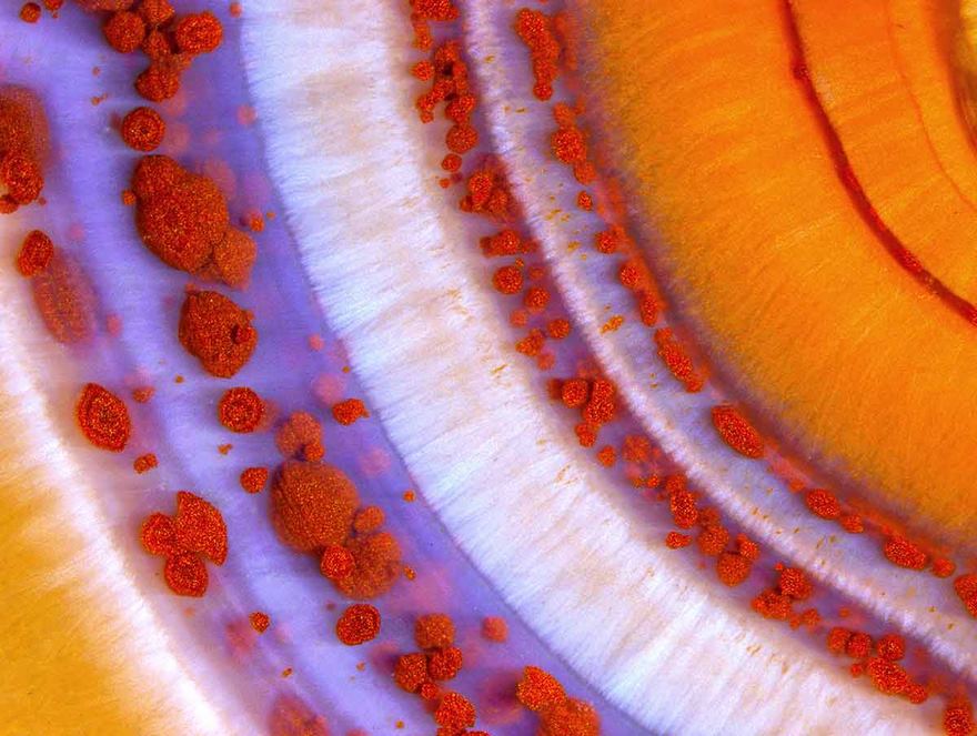
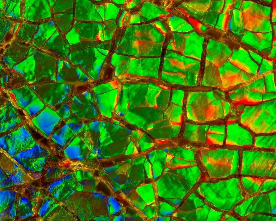
Second Place. Polished Slab Of Teepee Canyon Agate
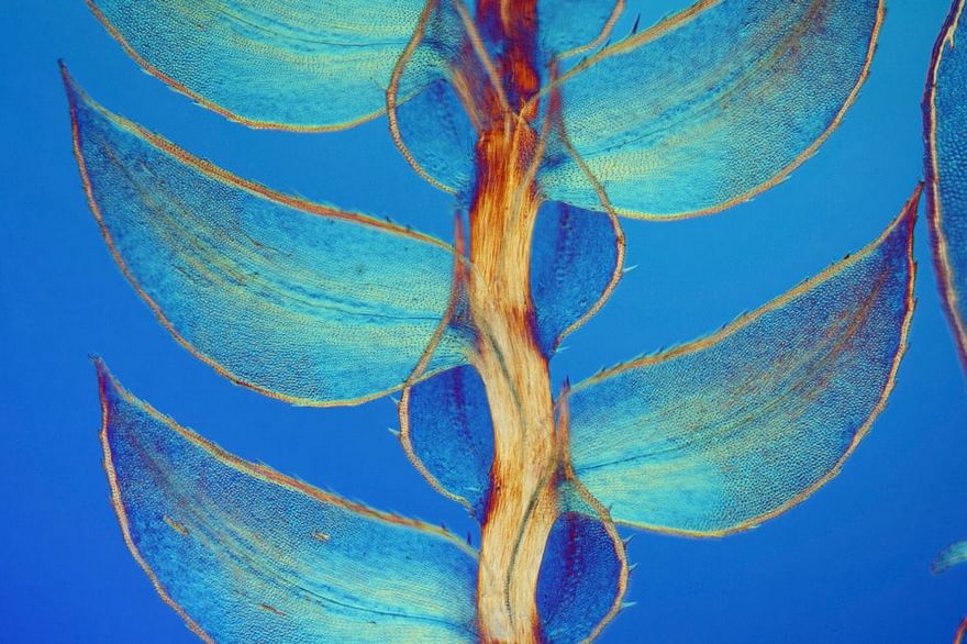
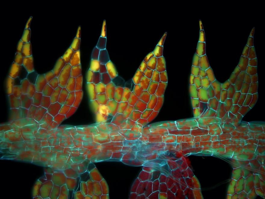
Seventh Place. Leaves Of Selaginella
Interesting. Because of the beautiful lighting, I would never have guessed this to be a fern.
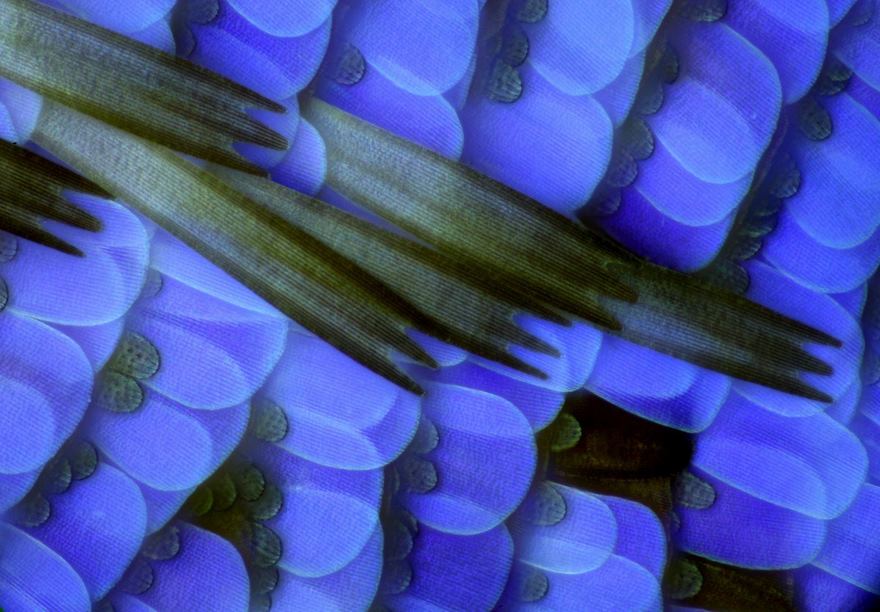
Scales Of A Butterfly Wing
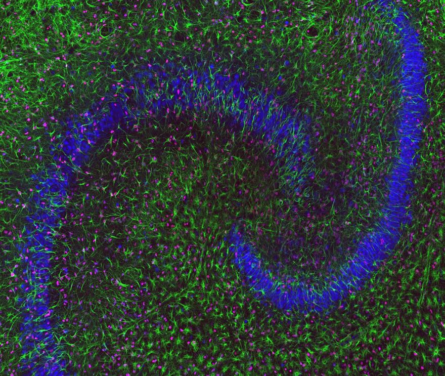
Hippocampal Neurons
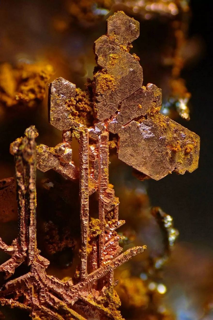
Copper Crystals
Scales Of A Butterfly Wing
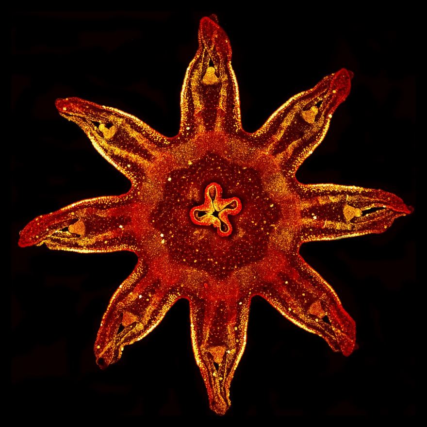
Jellyfish
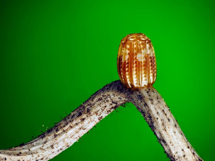
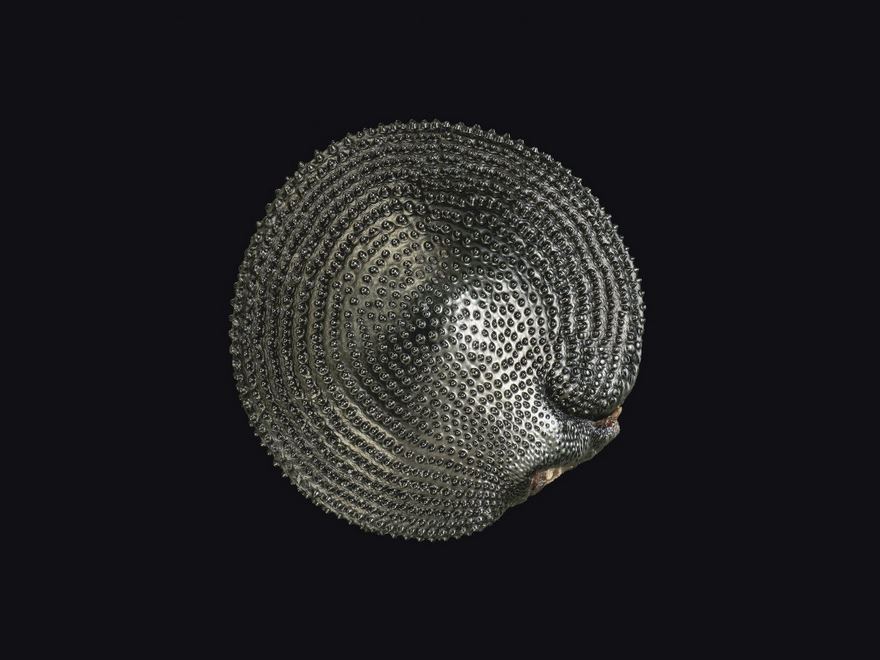
Egg Of A Gulf Fritillary Butterfly
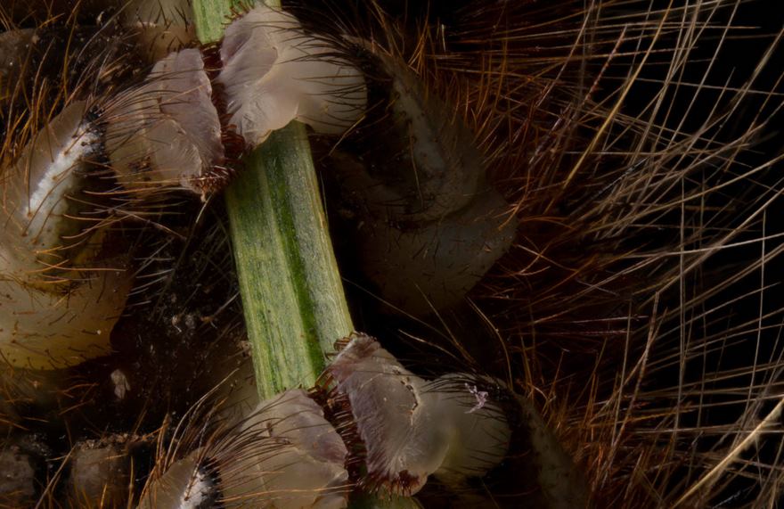
Prolegs Of A Hairy Caterpillar Gripping A Small Branch
Interference Patterns On A Glycerin Based Soapy Solution
This looks like an album cover. Hard-pressed to find a worthy bad though.
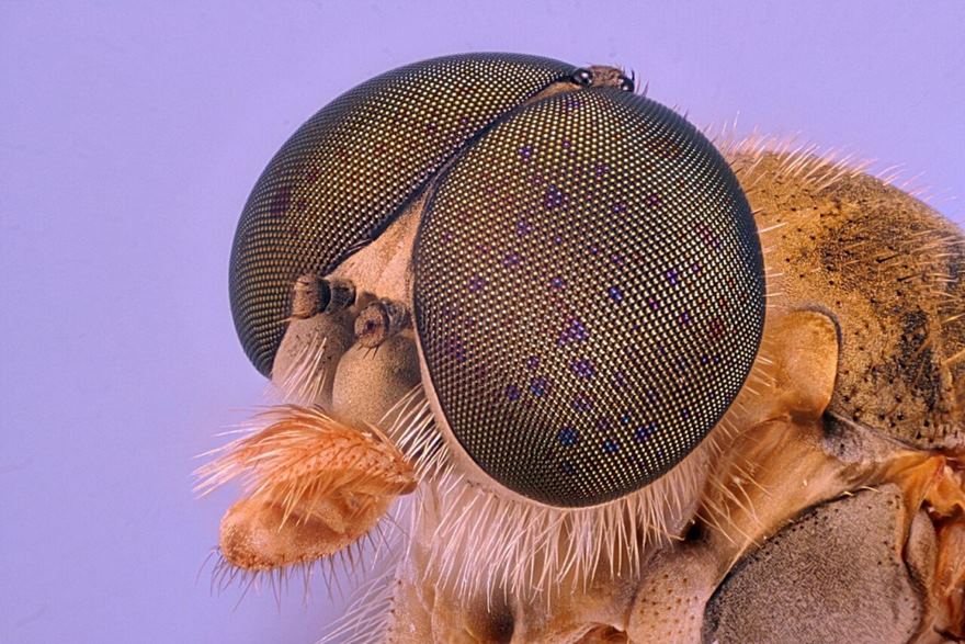
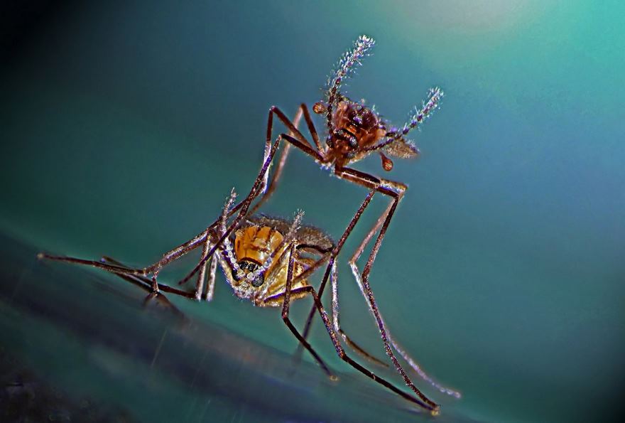
Robber Fly
Beta-Alanine And Taurine Crystals
Gears Coupling Hind Legs Of A Planthopper Nymph
Twentieth Place. Cow Dung
Quick Fact. Those liquid filled spores you see there are the fastest objects known to man(on earth). they accellerate faster than any bullet. This is so that the bacteria can land on a fresh piece of grass, far far away from the poo so that the cow can eat it again. Clever little spore right?
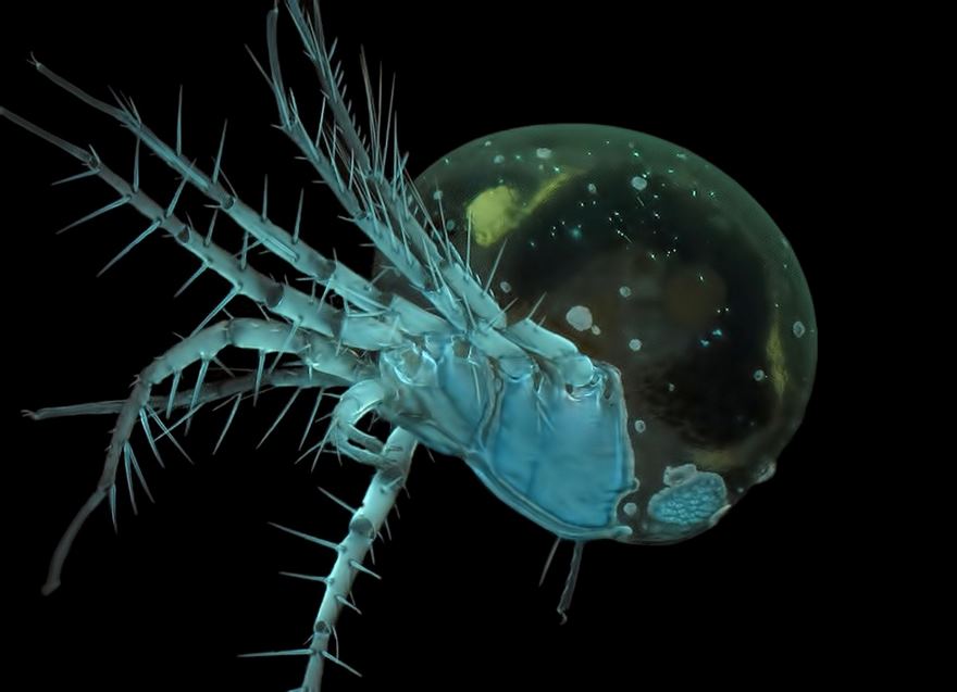
Water Mite
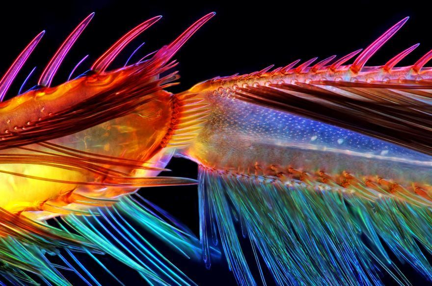
Leg Of A Water Boatman
Black Elder Tree Flower Stamen
Ant Leg
Wildflower Stamens
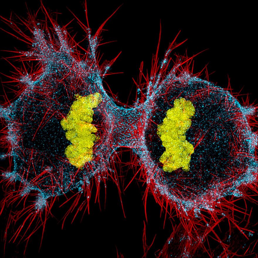
Twelfth Place. Human Hela Cell Undergoing Cell Division
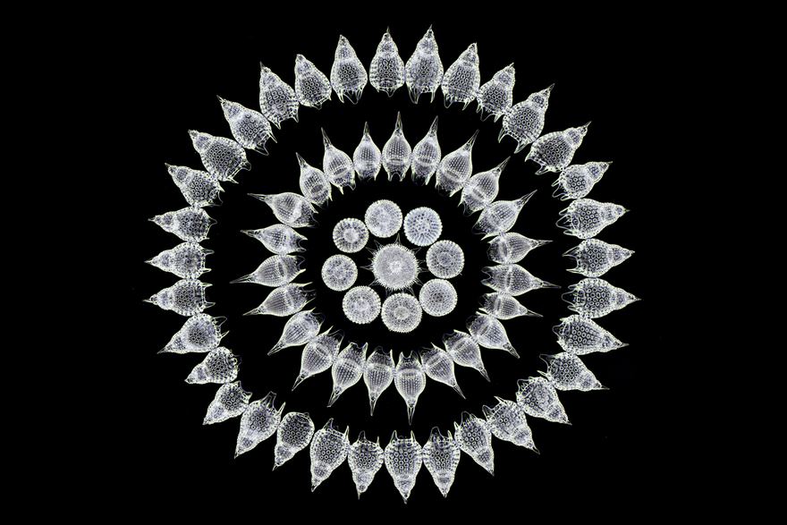
Sixteenth Place. 65 Fossil Radiolarians (Zooplankton) Carefully Arranged By Hand In Victorian Style
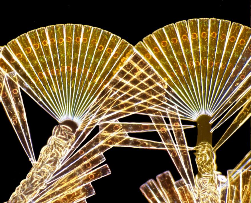
Licmophora Flabellata Diatoms
Third Place. Brain Cells From Skin Cells
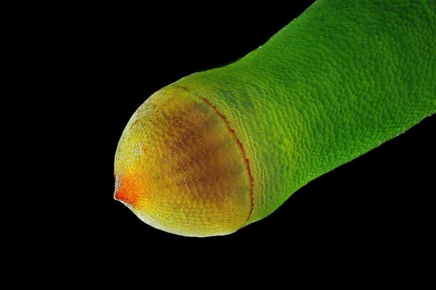
Spore Capsule Of A Moss
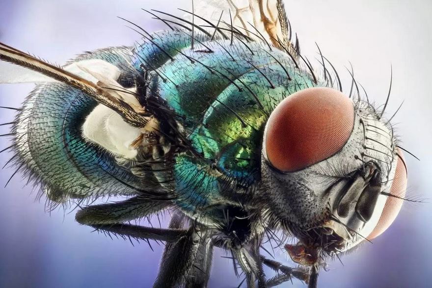
Green Bottle Fly
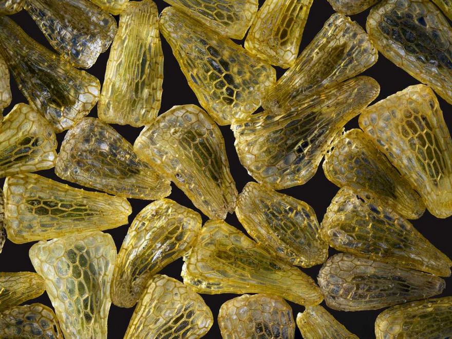
Curvepod Fumewort (Corydalis Curvisiliqua) Seed
Seeds Of An Indian Paintbrush Wildflower
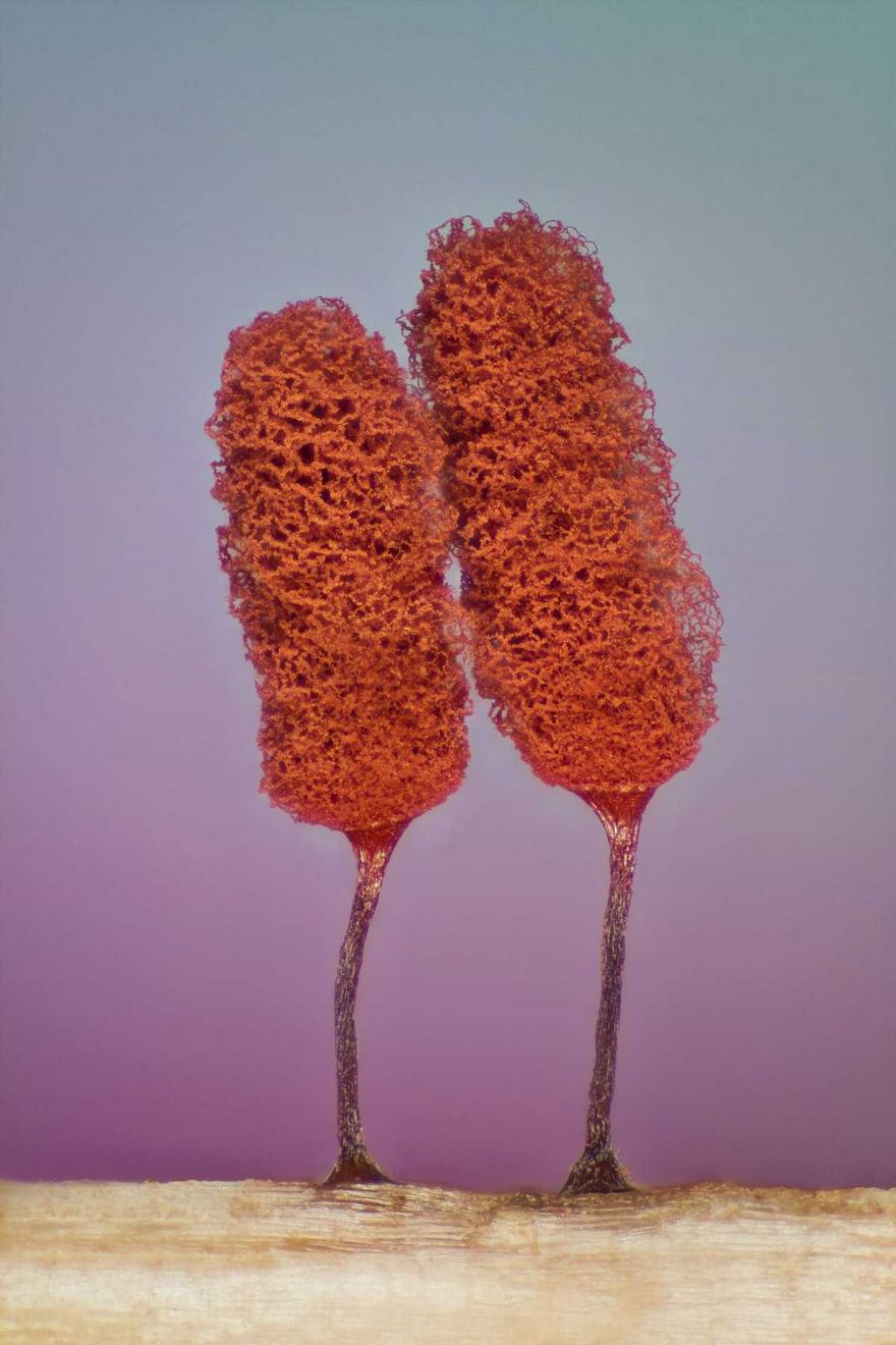
Slime Mold
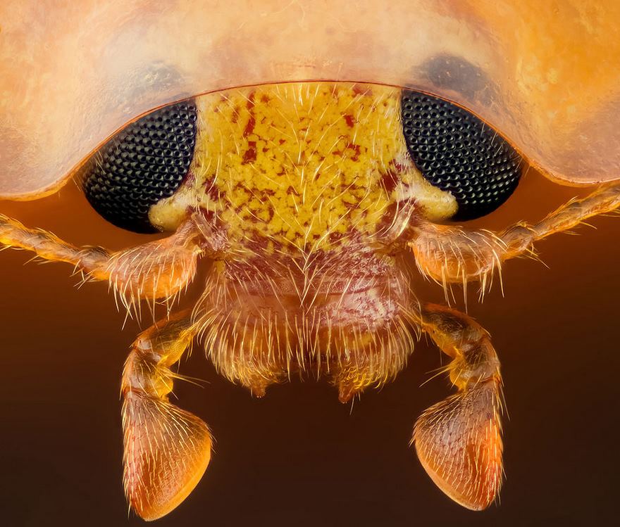
Fifteenth Place. Head Section Of An Orange Ladybird
For all of you future designers, great ideas for spaceship design.
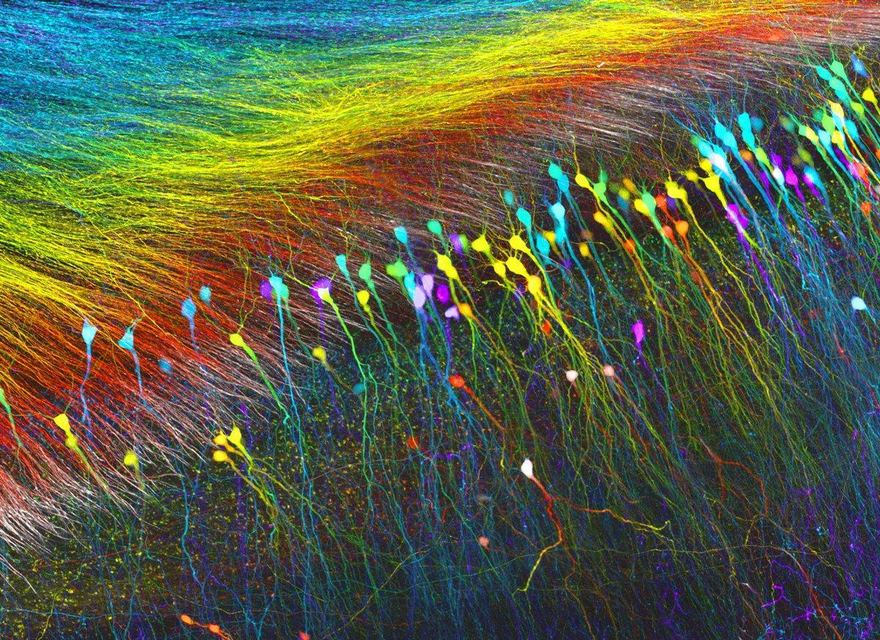
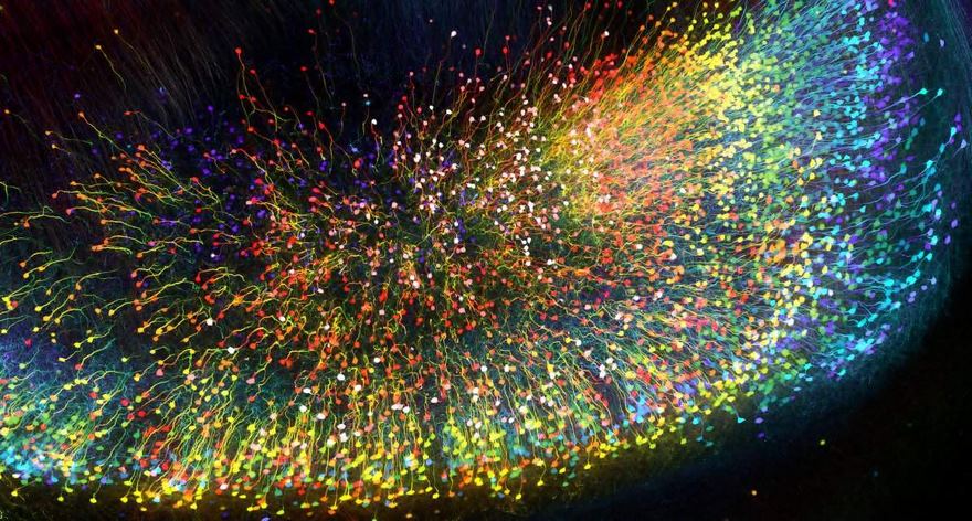
Dentate Gyrus Of A Optically-Cleared Transgenic Mouse Brain
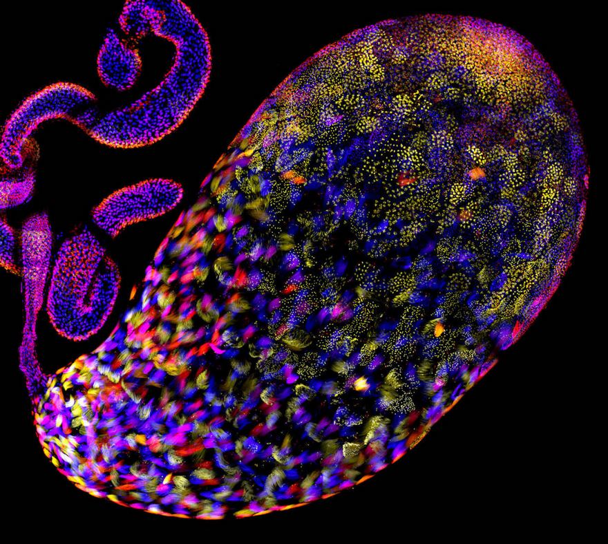
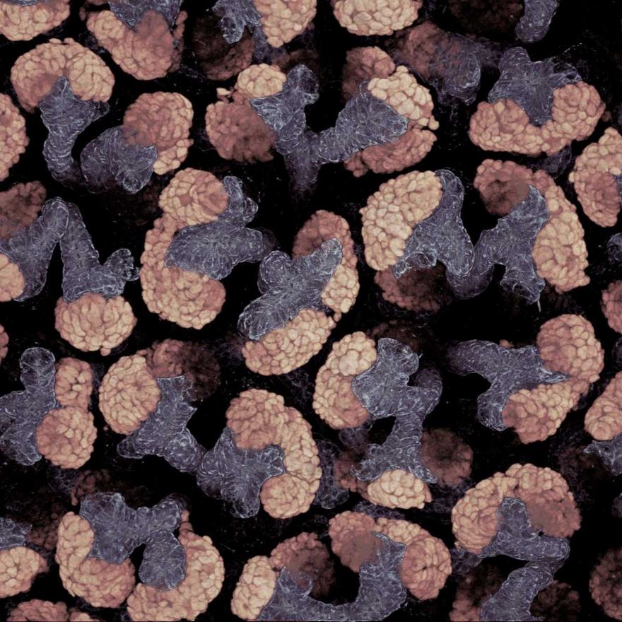
Testis Of A Fruit Fly
Algae
Microcrystal Test For Oxycodone Using Platinic Bromide Solution
Ammonite Shell
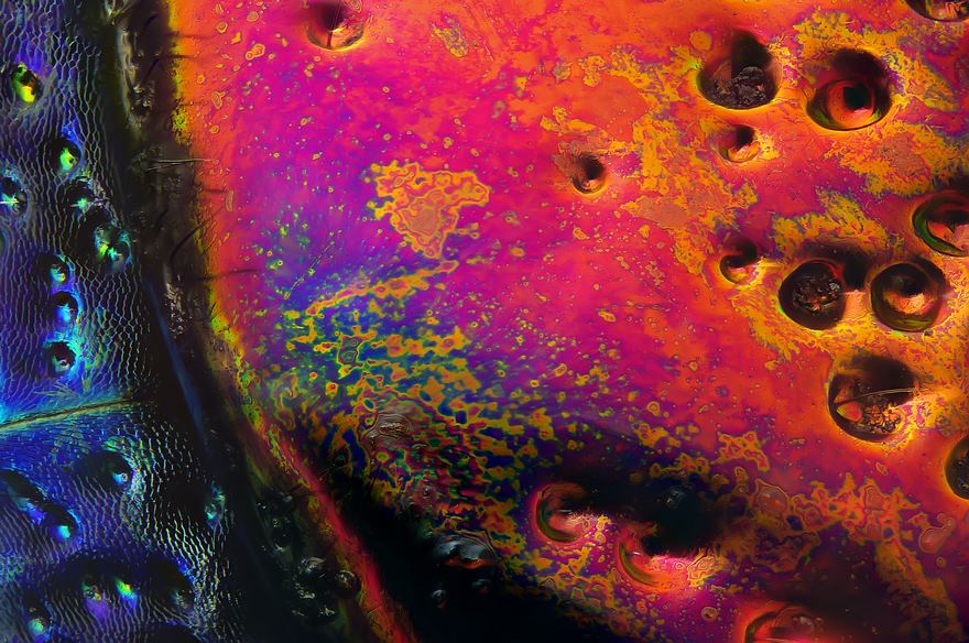
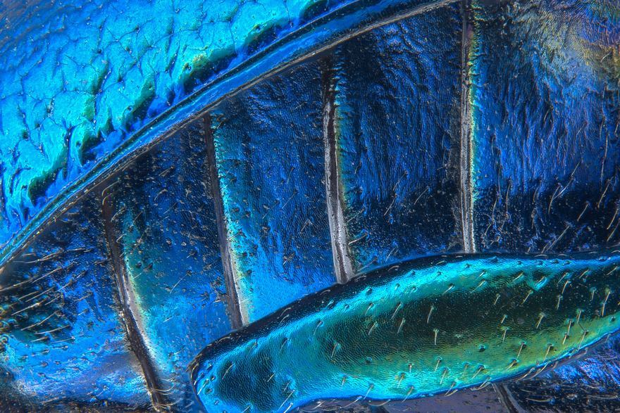
Section Of A Red Speckled Jewel Beetle
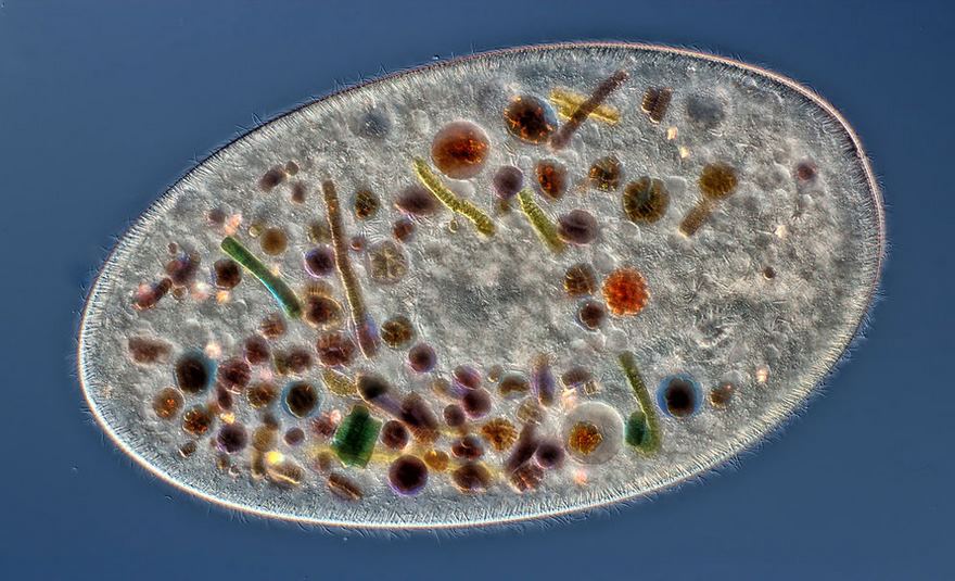
Tenth Place. Frontonia (Showing Ingested Food, Cilia, Mouth And Trichocysts)
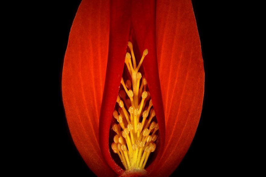
Section Of A Begonia Flower
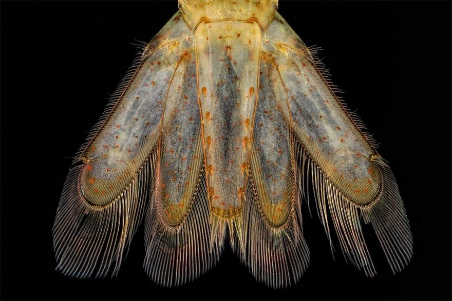
Tail Of A A Small Shrimp
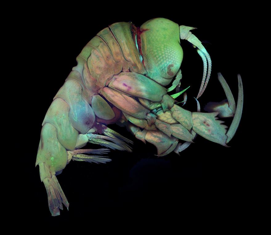
Deep Sea Crustacea
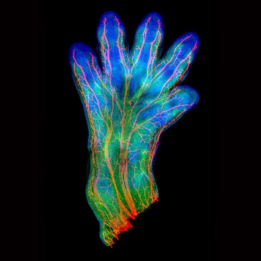
Mouse Hand, Showing Veins
How many pictures of mouse parts not attached have I seen eyes brains hands... sure multi coloured things are pretty but you didn't need to mutilate the poor creature
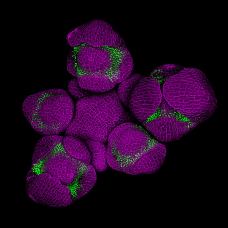
Young Flower Buds Of Arabidopsis
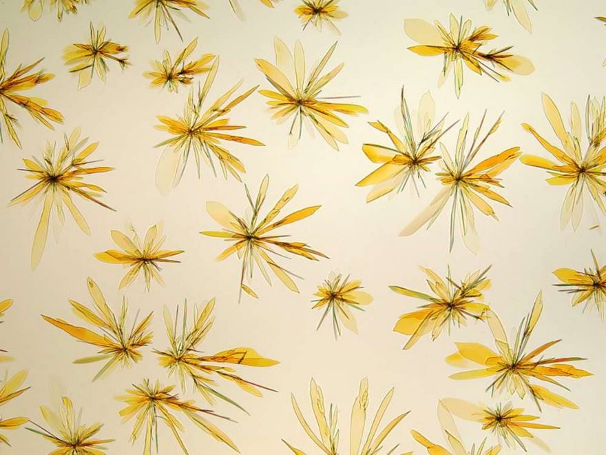
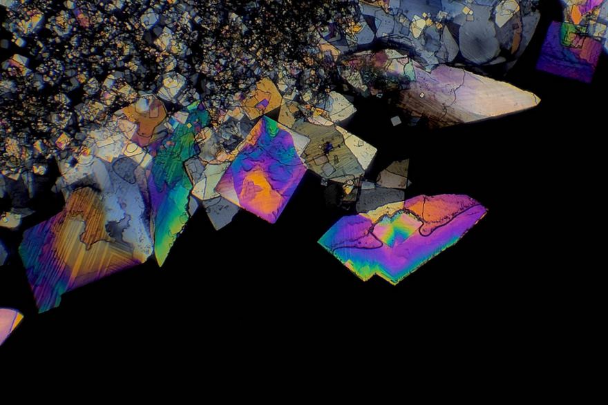
Diclofenac Crystals
Don't know what they are but I wish they were bigger it would make a good jewelry jewel
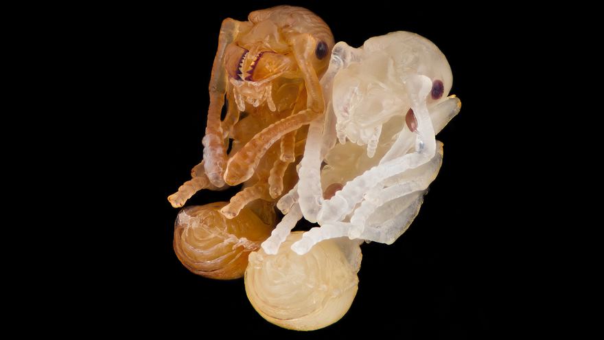
Ant Pupae
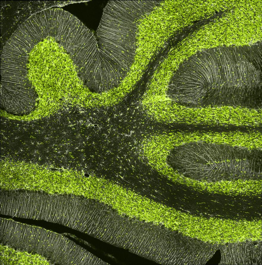
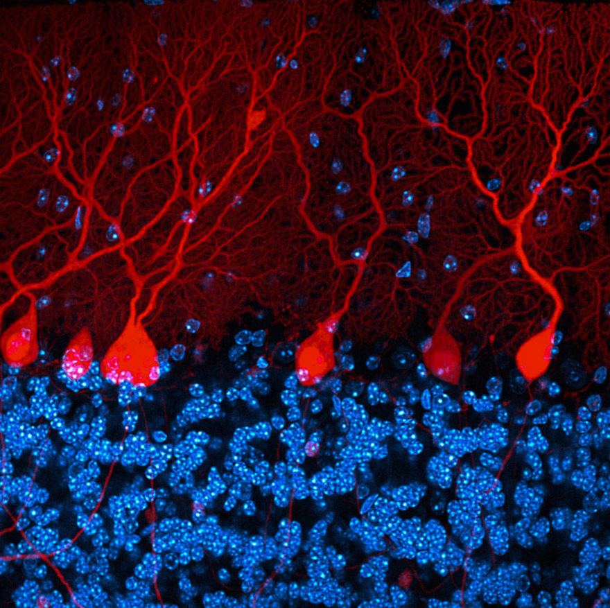
Cerebellum Brain Section Of A Rat
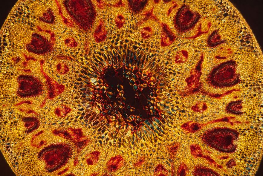
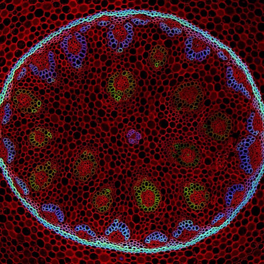
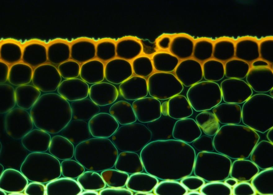
Cross Section Of Stem Of Barley
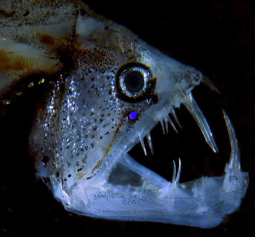
Viperfish
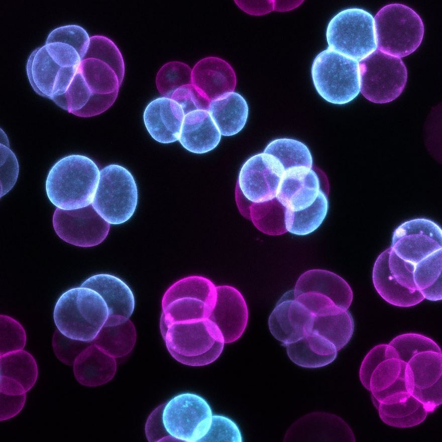
Early Stages Of Mouse Embryo Development
Fourteenth Place. Mouse Retinal Ganglion Cells
Section Of The Cerebellum
Galls Of A Mite
Surface Of Embryonic Mouse Kidney
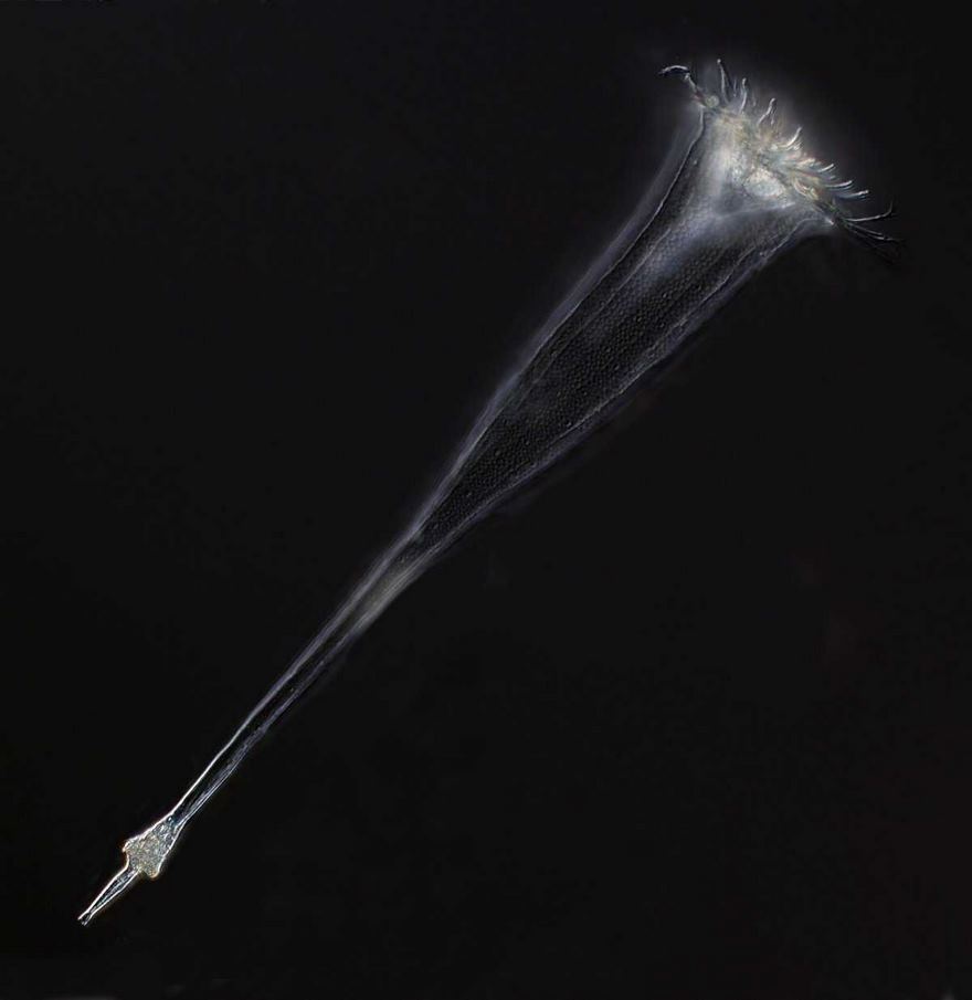
Tintinnid Ciliate Of The Marine Plankton
Hippocampal Slice Culture Stained For Neurons
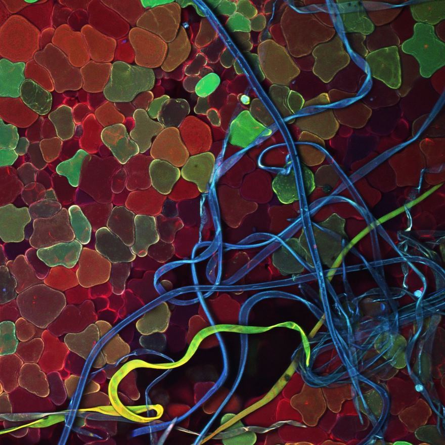
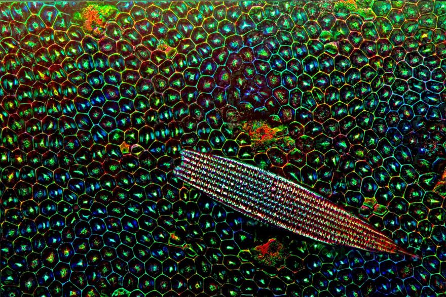
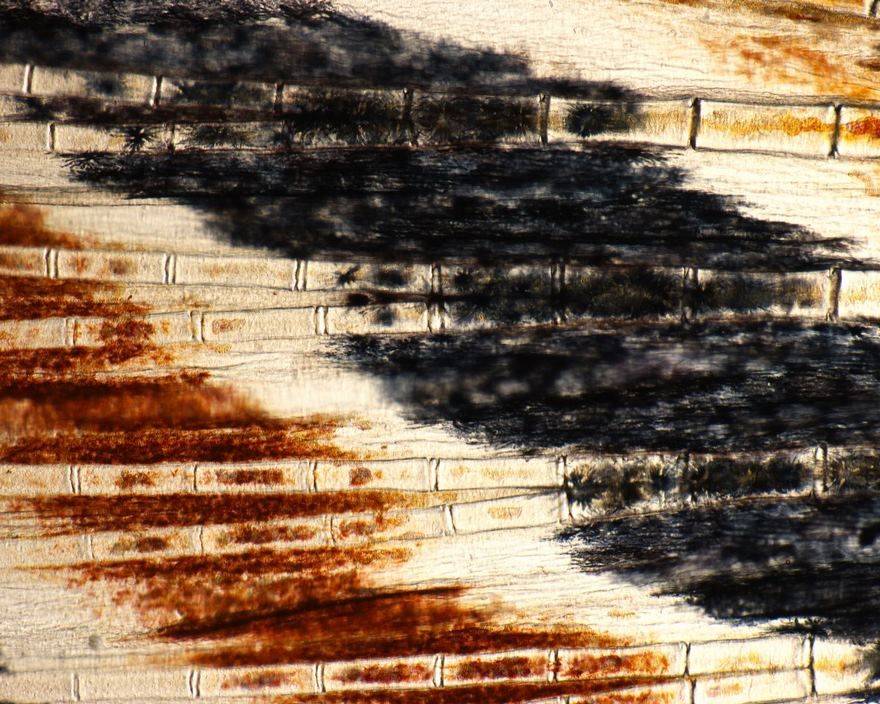
Forewing (Elytron) Of A Tiger Beetle
The forewing of the tiger beetle and the scale oft he forester moth are seen by the unaided human eye as a deep green. This picture with a magnification of 500x reveals that it is an additive color mixing of many color points originating from a combination of multilayer interference and dense arranged dimples in the insect cuticle.
Leaves Of A Liverwort
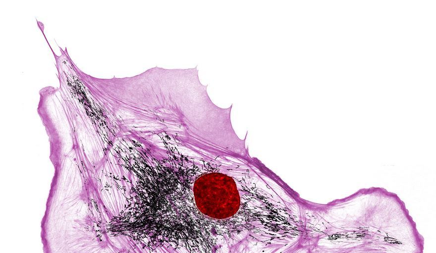
Actin, Mitochondria And Dna In A Bovine Pulmonary Artery Endothelial Cell
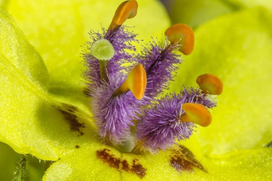
Mullein Flower
Eighteenth Place. Parts Of Wing-Cover
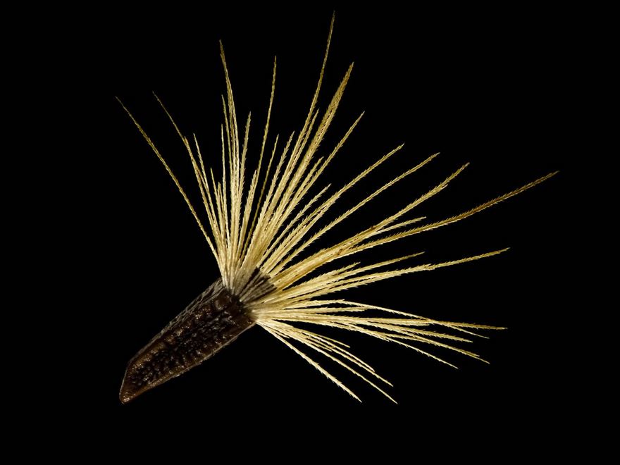
Jurinea Mollis Seed
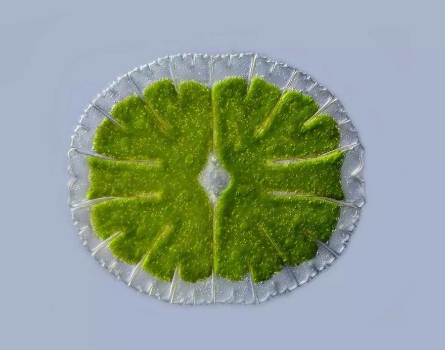
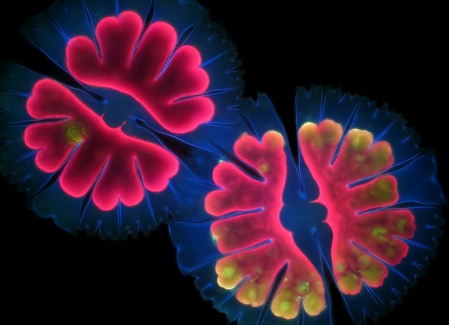
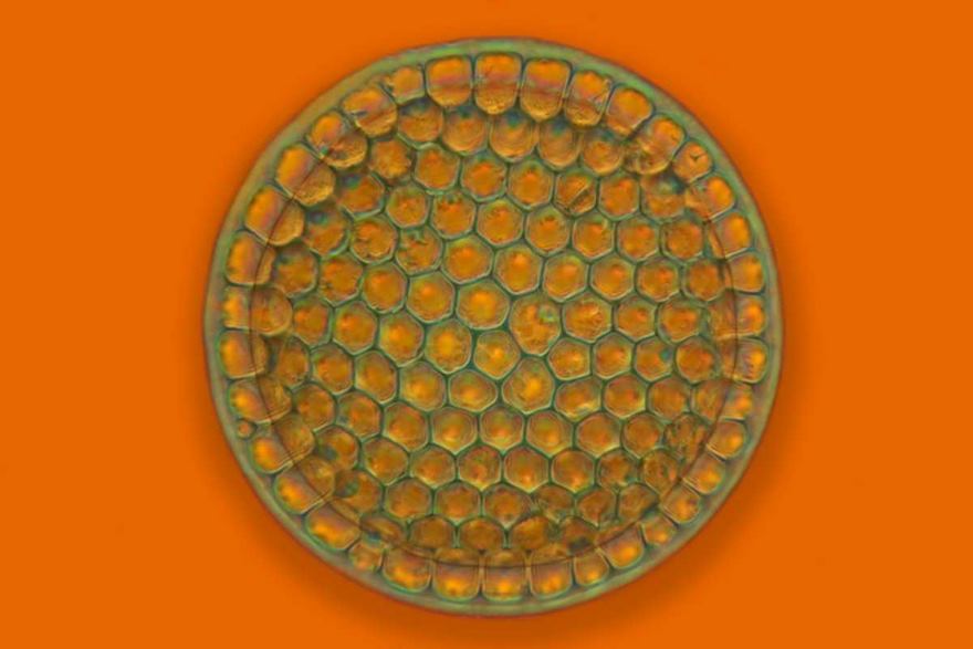
Micrasterias Thomasiana
Moving Vesicles
Cross Section Of A Lily Of The Valley
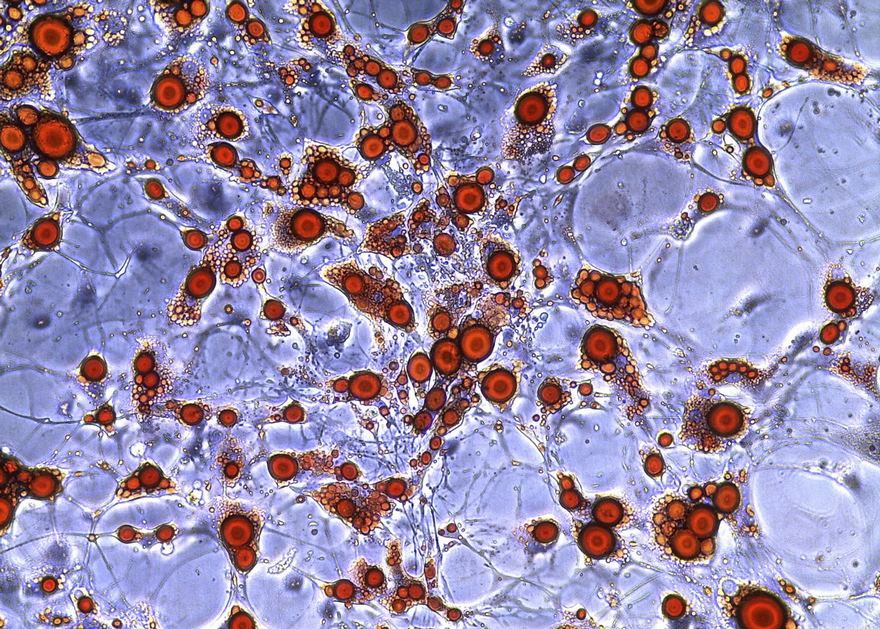
Cultured Fat Cells
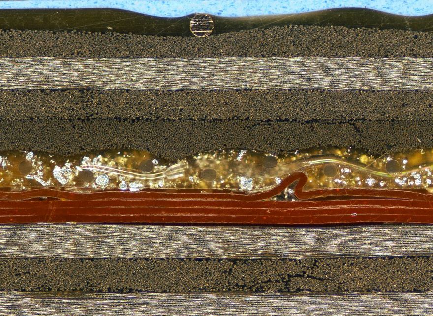
Cross-Section Through A Multi-Layered Carbon-Fiber Reinforced Composite Structure For Defect Analysis
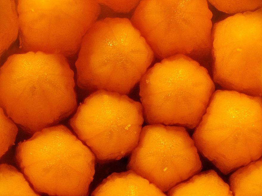
A Daisy’s Central Disc Pattern Of Tiny Unopened Flowers
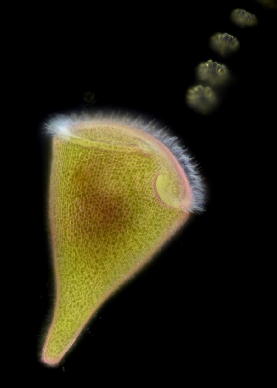
Trumpet Animalcule Containing Endosymbionts
Dasineura Affinis
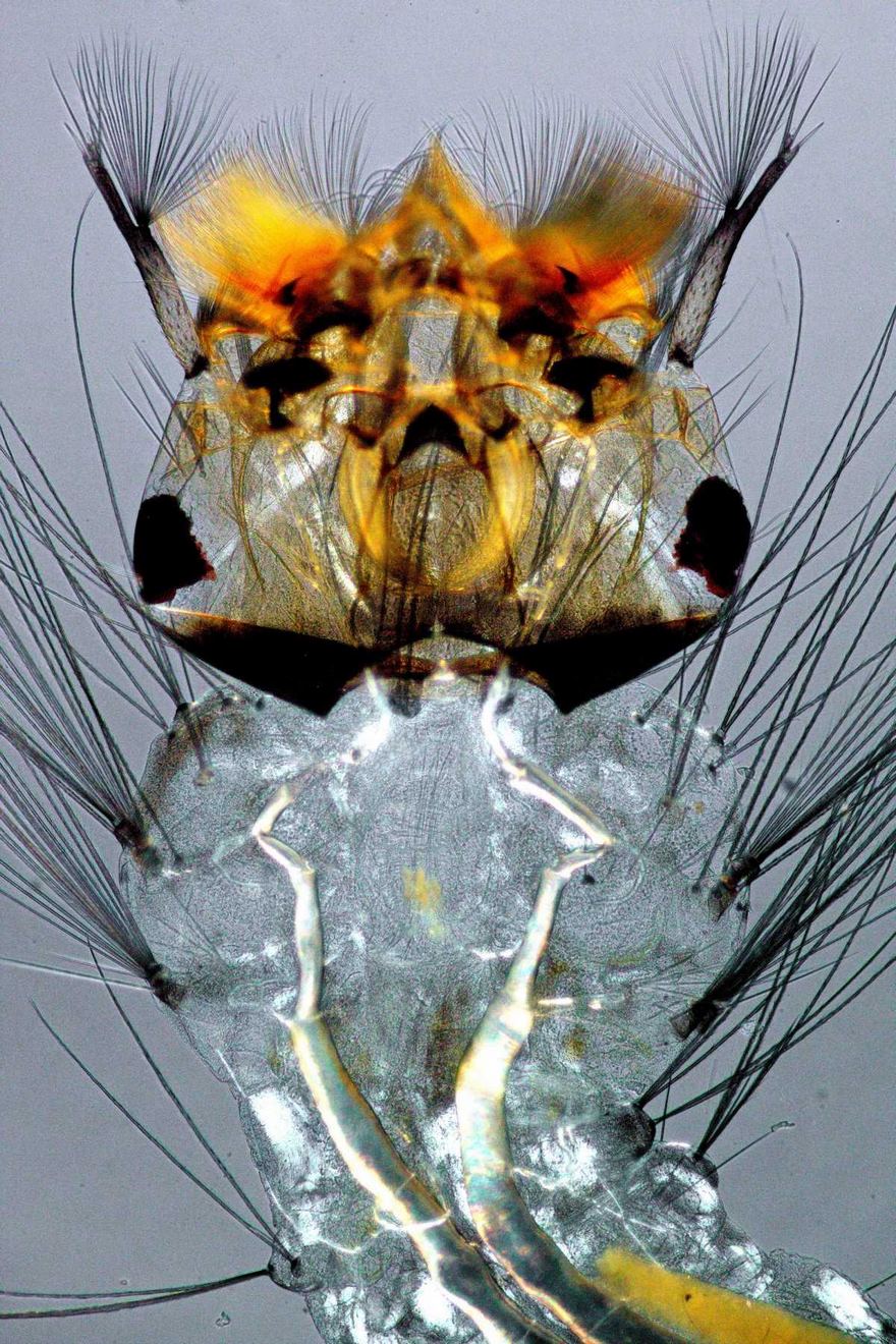
Mosquito Larva
Zebrafish Fin With Cylindrical Bone Segments And Rows Of Pigment
Fossil Diatom From Oamaru
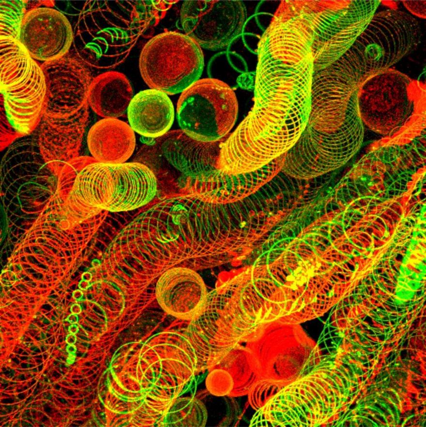
Section Of Stem Of A Bomarea Densiflora Plant Specimen
A few hundred years ago, biologists had to actually draw what they saw beyond the naked eye. Whether its 10X or 300X, the view under microscopes is mesmerizing.
A few hundred years ago, biologists had to actually draw what they saw beyond the naked eye. Whether its 10X or 300X, the view under microscopes is mesmerizing.

 Dark Mode

 No fees, cancel anytime


